Direct and indirect multiplex immunofluorescence (mIF) offer straightforward ways to explore spatial biology without specialized instruments. By combining multiple antibodies in the same sample, you can quickly visualize cell types, subcellular structures, and functional states to map how different proteins co-localize within tissue and how this architecture relates to disease biology.
Many low-plex IF experiments use either indirect detection with secondary antibodies or direct detection with fluorophore‑conjugated primaries—methods that can be performed on cells or tissue prepared in a variety of ways, depending on your experimental design. Both approaches fit into common IF workflows and standard widefield or confocal microscopes, but each has its own strengths and limitations for panel design, sample type, and future compatibility with higher-plex platforms.
Here, we cover two practical techniques for fluorescent staining using multiple antibodies in the same assay, and explain how newer tools like chimeric antibodies and the SignalStar® Multiplex IHC assay can take fluorescent multiplexing to the next level.
<< Jump to the Indirect IF Protocol Overview or the Direct IF Protocol Overview >>
Host-Based Multiplexing Using Indirect Immunofluorescence
Host‑based multiplexing is the standard approach for designing a low-plex IF experiment targeting 3–4 proteins within the same sample. With this method, each primary antibody is raised in a different host species (for example, rabbit and mouse), and species‑specific secondary antibodies—each coupled to a distinct fluorophore—are used to detect them. This is referred to as indirect detection because the fluorescent signal comes from the secondary antibody, not the primary.
 |
 |
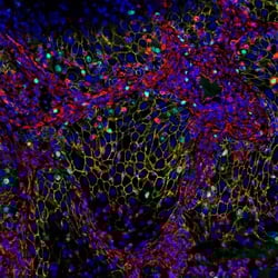 |
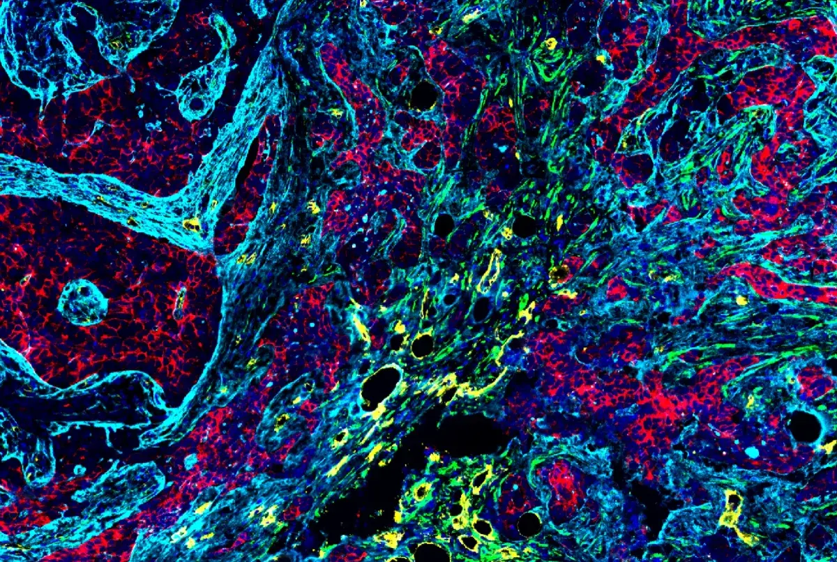
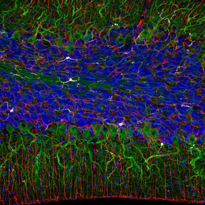
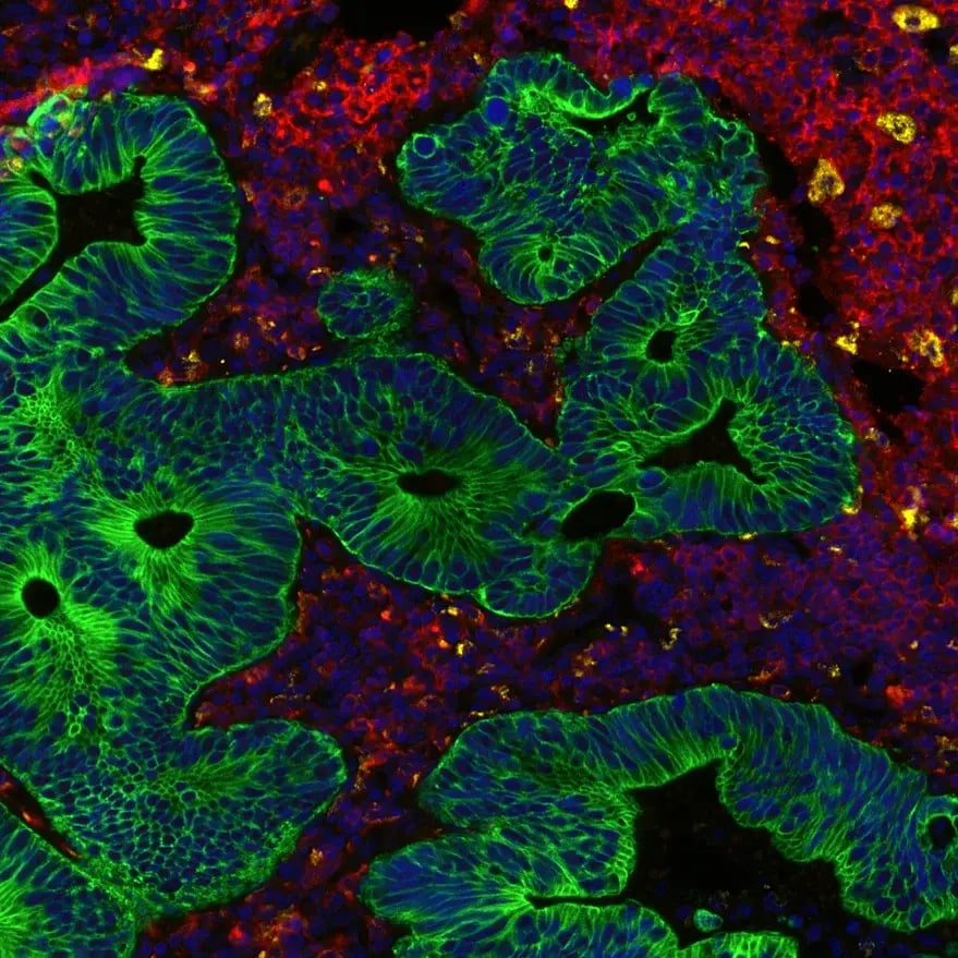
Host-based multiplexing using indirect immunofluorescence in cells, frozen tissue, and paraffin-embedded tissue (IHC-Paraffin). Left: HCT 116 cells treated with Chloroquine #14774 (50 µM, overnight) using LC3B (E5Q2K) Mouse Monoclonal Antibody #83506 (green), beta-Actin (13E5) Rabbit Monoclonal Antibody #4970, and DAPI #4083 (blue).
Center: Fixed frozen mouse cerebellum using β3-Tubulin (D71G9) Horse Chimeric Monoclonal Antibody #59820 (green), Iba1/AIF-1 (E4O4W) Feline Chimeric Monoclonal Antibody #87276 (gray), GFAP (E4L7M) Rabbit Monoclonal Antibody #80788 (red), and DAPI #4083 (blue). Rabbit primary antibody is detected using an anti-rabbit Fc-specific secondary antibody.
Right: Paraffin-embedded human squamous cell lung carcinoma using KI-67 (D3B5) Feline Chimeric Monoclonal Antibody #43021 (green), beta-Catenin Mouse Chimeric Monoclonal Antibody (yellow), Vimentin (D21H3) Rabbit Monoclonal Antibody #5741 (red), and DAPI #4083 (blue). Rabbit primary antibody is detected using an anti-rabbit Fc-specific secondary antibody.
Why use host‑based multiplexing?
One of the main benefits of host‑based multiplexing is that it provides a brighter signal than most direct IF methods because multiple fluorophores can bind to each primary via the secondary antibody, amplifying the signal. This method also adds some freedom in experimental design, as you can experiment with which fluorophore to pair with which primary. The trade‑offs are the need for high-quality secondary antibodies to avoid cross‑reactivity and background signal, as well as the extra secondary incubation step.
Host‑based indirect IF is a great entry point into multiplex imaging for cultured cells, fixed frozen tissue sections, and many FFPE tissues, especially when you want to visualize 2–4 markers on a fluorescent microscope already in your lab. Panels built this way are often conceptually compatible with higher‑content multiplex spatial biology platforms that use secondary antibodies or similar indirect detection schemes, making it easier to scale up your biology later on platforms such as the COMET System by Lunaphore or other cyclic immunofluorescence platforms.
Using Chimeric Antibodies to Expand Host‑Based Multiplexing
Historically, host‑based multiplexing has been limited by the availability and quality of non‑rabbit primary antibodies. CST chimeric antibodies help solve this limitation by taking the binding domain of a well‑validated rabbit monoclonal and engineering it onto a different host backbone. Each chimeric antibody keeps the same specificity as the original rabbit clone, but can now be detected with a distinct species‑specific secondary, unlocking more channels within the same experiment.
Blog: Anything but Rabbit: Why Multiplex with Chimeric Antibodies?
Because CST chimerics are designed to work within standard IF and IHC workflows and are validated for use in fixed frozen and FFPE tissues, you can add them to existing host‑based multiplex panels without changing your assay conditions or imaging setup. This flexibility allows you to build complex panels using antibodies that are proven to be specific and sensitive, making it valuable for studies in neuroscience, immunology, and other fields that rely on multiplex imaging.

Indirect IF Protocol Overview (Host‑Based Multiplexing)
Below is a general protocol overview for indirect IF using antibodies raised in different host species (for example, rabbit and mouse) and/or chimeric antibodies from alternative host species. Note that the actual protocol will vary based on the specific products’ recommended protocol.
-
Sample Preparation: Follow the suggested protocol listed on the product webpage or datasheet for all CST antibodies. Some optimization and additional testing may be required.
- Antigen Unmasking (!! FFPE tissue only !!): Heat slides submersed in 1X EDTA unmasking solution in a microwave until boiling; follow with 15 min at sub-boiling temperature (95°-98°C). No cooling is necessary.
-
Rinse: Briefly rinse with 1X PBS.
-
Block: Block with a blocking buffer for 60 min to avoid non-specific binding of the secondary antibody.
-
NOTE: This step is especially important when using multiple species‑specific secondaries in the same sample.
-
-
Dilute & Incubate Primaries: Dilute each primary antibody to its recommended dilution as indicated on the product webpage or datasheet using antibody dilution buffer; aspirate blocking solution, then apply diluted primary antibody. Incubate overnight at 4°C.
-
Wash: Wash 3 times with 1X PBS for 5 min each.
-
Dilute & Incubate Secondaries: Dilute each fluorophore-conjugated secondary antibody to its recommended dilution as indicated on the product webpage or datasheet using an antibody dilution buffer, then apply diluted secondary antibody. Incubate for 1 hr at room temperature
-
NOTE: Choose secondaries that are species‑specific (for example, anti‑rabbit Fc, anti‑mouse, anti‑horse, anti‑feline) and conjugated to fluorophores with minimal spectral overlap. When using chimeric antibodies, it is crucial to use an anti-rabbit Fc-specific antibody instead of any anti-rabbit secondary.
-
-
Counterstain: Perform any counterstaining steps using complementary reagents (such as DAPI), mount in antifade reagent.
When you are designing panels for frozen tissue, use antibodies that have been validated for IF‑frozen and confirmed to work in the relevant species, since fixation and antigen retrieval requirements differ from FFPE. Similar host‑based multiplexing strategies can be adapted for FFPE tissue, provided you use FFPE‑validated antibodies and appropriate antigen retrieval conditions.
Direct IF with Fluorophore‑Conjugated Primary Antibodies
Direct IF uses primary antibodies conjugated to fluorophores with different spectra, eliminating the need for secondary antibodies. This remains a powerful multiplexing strategy, particularly for 2–3 markers, and can be an efficient and effective way to get started with multiplex imaging.
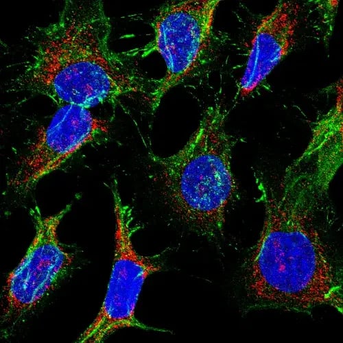
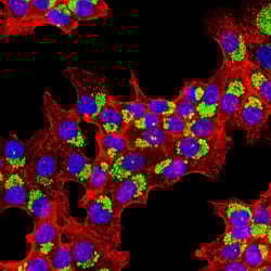
Direct IF using fluorophore-conjugated primary antibodies in cells, frozen tissue, and paraffin-embedded tissue (IHC-Paraffin). Left: HeLa cells using β-Catenin (D10A8) Rabbit Monoclonal Antibody (Alexa Fluor® 488 Conjugate) #88187 (green), MTHFD2 (E7A4L) Rabbit Monoclonal Antibody (Alexa Fluor® 647 Conjugate) #64107 (red), and DAPI #4083 (blue).
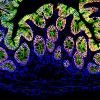
Center: Fixed frozen mouse colon. Sections were labeled with E-Cadherin (24E10) Rabbit Monoclonal Antibody #3195 (red) prior to blocking free secondary binding sites with Rabbit (DA1E) Monoclonal Antibody IgG Isotype Control #3900. Sections were then labeled with β-Catenin (D10A8) Rabbit Monoclonal Antibody (Alexa Fluor® 488 Conjugate) #88187 (green) and DAPI #4083 (blue).
Right: Paraffin-embedded human colon adenocarcinoma using HLA-DRA (E9R2Q) Rabbit Monoclonal Antibody #97971 (Alexa Fluor® 750 Conjugate) (red), CD68 (D4B9C) Rabbit Monoclonal Antibody (Alexa Fluor® 488 Conjugate) #24850 (yellow), Pan-Keratin (Type I) (E6S1S) Rabbit Monoclonal Antibody (Alexa Fluor® 647 Conjugate) #75435 (green) and DAPI #4083 (blue).
Why use direct IF multiplexing?
Direct IF multiplexing is well‑suited when you want a streamlined protocol, without a secondary antibody step, which shortens hands‑on time and reduces opportunities for pipetting errors or cross‑reactive secondaries. By using multiple antibodies from the same host species (often rabbit) in one experiment, you don’t have to worry about species‑specific secondary selection, which simplifies panel design
%20Bain%2c%20PhD%20CST%20Immunofluorescence.png?width=175&height=175&name=Virginia%20(Ginny)%20Bain%2c%20PhD%20CST%20Immunofluorescence.png)
Virginia (Ginny) Bain |
“Direct IF with conjugates is a very powerful tool—it allows you to multiplex your favorite antibodies from the same species without worrying about cross-reactivity. This enables you to use antibodies that have already proven successful in a new, effective way. It's a simple and elegant solution for assay design." |
Because the signal comes from individual fluorophore‑conjugated primary antibodies, direct IF typically produces a lower signal than indirect IF but can still be more than sufficient for moderately or highly expressed targets. Direct IF works well on standard widefield or confocal microscopes and is compatible with both cultured cells and tissue sections, provided the antibodies are validated for IF in the relevant sample type (fixed frozen or FFPE), and the chosen fluorophores match your filter sets. These panels may also be compatible with common imaging platforms, such as the Cell DIVE Multiplex Imaging Solution by Leica Microsystems, depending on the fluorophores used.
When should I use direct vs indirect IF?
-
Use indirect, host‑based IF when you need higher sensitivity for low‑abundance targets, want to use the same primary antibody with different secondaries, or plan to expand into host‑based multiplexing with chimerics and secondary detection on tissue.
-
Use direct IF when your targets are relatively abundant, you prefer a faster protocol, you need to avoid mouse-on-mouse background, or you want to combine several rabbit antibodies in one experiment.
In practice, many labs combine these strategies—for example, using indirect detection for dim targets and direct conjugates for brighter markers in the same sample—so long as fluorophore selection and imaging settings are carefully planned. You can learn more about designing complex panels that combine direct and indirect IF in this blog: Combining Direct and Indirect Multiplex IF: Strategies with Conjugated Antibodies.
Direct IF Protocol Using Antibodies Conjugated to Different Fluorophores
Below is a general protocol overview for direct IF staining using primary antibodies directly conjugated to different fluorophores. As always, consult individual product datasheets for specific recommendations.
-
Sample Preparation: Follow the suggested protocol listed on the product webpage or datasheet for all CST antibodies. Some optimization and additional testing may be required.
- Antigen Unmasking (!! FFPE tissue only !!): Heat slides submersed in 1X EDTA unmasking solution in a microwave until boiling; follow with 15 min at sub-boiling temperature (95°-98°C). No cooling is necessary.
-
Rinse: Briefly rinse with 1X PBS.
-
Block: Block with a blocking buffer for 60 min.
-
Dilute & Incubate: Dilute each primary antibody to its recommended dilution as indicated on the product webpage or datasheet using antibody dilution buffer; aspirate blocking solution then apply diluted primary antibody. Incubate overnight at 4°C.
-
Wash: Wash 3 times with 1X PBS for 5 min each.
-
Counterstain: Perform any counterstaining steps using complementary reagents (such as DAPI), mount in antifade reagent.
When designing direct IF panels, choose fluorophores with separated excitation and emission spectra that are compatible with your microscope and avoid spectral overlap. CST offers a range of directly conjugated monoclonal antibodies covering common colors, making it straightforward to assemble 2–4-color panels for basic multiplex experiments.
Putting It All Together: Next Steps in Multiplex Spatial Biology
Indirect and direct multiplex IF are the most accessible ways to start exploring multiplex tissue imaging and spatial biology using existing microscopes and standard workflows. By carefully choosing between host‑based indirect IF, direct IF, or a combination of both—and leveraging chimeric antibodies when you need more hosts—you can build low‑plex panels that answer meaningful biological questions without specialized instruments.
As your questions become more complex and you want to measure more markers or lower‑abundance functional targets in FFPE tissue, you may need to move beyond low‑plex IF into mid‑plex spatial biology platforms. The SignalStar Multiplex IHC assay offers a natural next step: The technology uses oligonucleotide‑based amplification to detect multiple phenotypic and functional markers simultaneously in FFPE sections, enabling up to 8‑plex amplified spatial profiling in a 2‑day, non‑cyclic workflow.
Whether you are designing your first three‑color IF experiment or planning a transition toward higher‑plex spatial biology, selecting application‑validated antibodies, matching them to your sample type and imaging setup, and planning for future platform compatibility will help ensure reliable, biologically meaningful data. CST resources on immunofluorescence, chimeric antibodies, and multiplex spatial biology can guide you at each step—from panel design and protocol optimization to the move from low‑plex microscopy to mid‑plex spatial platforms like the SignalStar technology and beyond.