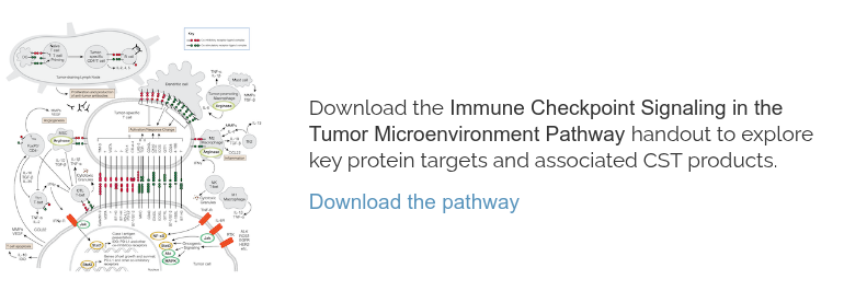
This blog explores the mechanisms, pathways, and proteins involved in one of the Hallmarks of Cancer—Avoiding Immune Destruction. Identified as a hallmark in 2011, ten years after the seminal “The Hallmarks of Cancer” paper was published in Cell, the concept reflects the critical role the immune system plays in both suppressing and, when evaded by tumor cells, enabling cancer progression.
Recognizing this hallmark has helped advance cancer research and therapy, leading to the rise of immunotherapies that harness the body’s natural defenses to target cancer cells.
< Jump to the product list at the end of this blog >
How does the immune system attack cancer?
The immune system detects and eliminates cancer cells in a multistep process that involves many different types of immune cells. Among the best-known players in cancer immune defense are cytotoxic T cells, identified by the surface expression of CD8, which serves as a co-receptor for the T cell receptor (TCR).
|
When a CD8+ T cell detects a cancer cell via the recognition of peptide antigens presented by major histocompatibility complex (MHC) class I, the resulting signaling cascade triggers the release of lysosome-like granules. These contain perforin, a membrane pore-forming protein, and granzymes such as Granzyme B, which enter the cancer cell and activate caspases to induce programmed cell death via apoptosis. |
|
What are the Hallmarks of Cancer?The Hallmarks of Cancer1-3 are a research framework that organizes the key traits cancer cells acquire in order to grow and spread. Initially described by Douglas Hanahan and Robert Weinberg in 2000, the framework groups the underlying mechanisms of cancer into a series of smaller subsets to advance discovery. The concept was expanded in 2011 to include two additional hallmarks and two enabling characteristics, and again in 2022 with four new emerging hallmarks.
|
|
This highly efficient mechanism enables CD8+ T cells to identify and kill target cells while minimizing damage to surrounding healthy tissue.
 |
 |
|
Immunohistochemical (IHC) analysis of paraffin-embedded human lung carcinoma tissue using CD8α(D8A8Y) Rabbit mAb #85336, highlighting the presence of cytotoxic CD8+ T cells—key immune cells involved in the detection and elimination of cancer cells.
|
IHC analysis of paraffin-embedded human B-cell non-Hodgkin lymphoma using Granzyme B (D6E9W) Rabbit mAb (Alexa Fluor® 488 Conjugate) #49060 (green) and DAPI #4083 (blue), demonstrating the presence of Granzyme B in tumor-infiltrating lymphocytes, a protein indicating cytotoxic potential and a key player in a mechanism by which cytotoxic T cells mediate cancer cell apoptosis after antigen recognition. |
However, cancer cells have evolved many mechanisms to evade T cell-mediated immune destruction, some of which are discussed below.
The Evasion Equation: How do cancer cells evade the immune system?
Immune Exclusion
To successfully target cancer cells, CD8+ T cells—and the immune cells required for their activation, such as dendritic cells—must be able to penetrate solid tumors. However, many tumors can prevent the infiltration of anti-tumor immune cells through a process known as immune exclusion.
Immune exclusion is achieved through several mechanisms:
- Mechanical Barriers: Cancer-associated fibroblasts (CAFs) within the tumor microenvironment (TME) promote collagen production and fibrosis to form a dense stromal barrier that physically blocks immune cells. These fibroblasts can be identified by markers including fibroblast activation protein (FAP) and leucine-rich repeat-containing protein 15 (LRRC15).

IHC analysis of paraffin-embedded human urothelial carcinoma using FAP (F1A4G) Rabbit mAb (Alexa Fluor® 647 Conjugate) #64706 (red) and DAPI #4083 (blue), highlighting CAFs within the TME, which contribute to the formation of stromal barriers that block immune cell infiltration. - Suppressed Chemokine Expression: Tumors can also reduce the expression of chemokines, including CXCL9, CXCL10, and CCL5, that would normally recruit immune cells to the TME. This suppression can occur either through genetic alterations, the downregulation of pathways that lead to chemokine production, or through the exclusion of chemokine-producing immune cells.


IHC analysis of paraffin-embedded non-small cell lung cancer using CXCL9/MIG (E6Z5W) Rabbit mAb #30327. IHC analysis of paraffin-embedded human prostate cancer using CXCL10 (F9N8I) Rabbit mAb #45008. IHC analysis of paraffin-embedded human non-small cell lung cancer using CCL5/RANTES (E9S2K) Rabbit mAb #36467.
- CAF-Derived Chemokines: CAFs can also prevent CD8+ T cell infiltration by producing the chemokine CXCL12, which leads CD8+ T cells away from the TME to further reduce their infiltration. Consequently, CXCL12 is of interest as a potential therapeutic target.

IHC analysis of paraffin-embedded human prostate carcinoma using SDF1/CXCL12 (D8G6H) Rabbit mAb #97958. The analysis shows that CXCL12 is present and therefore may be contributing to the tumor's ability to evade immune destruction.
Defects in Antigen Presentation
If CD8+ T cells manage to successfully infiltrate the TME, an effective immune response depends on their ability to recognize tumor cells via the presentation of cancer-associated or cancer-specific peptide antigens by MHC class I molecules.
Cancer cells can evade detection by downregulating antigen presentation. This immune escape can occur through downregulation of gene expression or mutations in the antigen presentation machinery, including human leukocyte antigen (HLA) class I and β2-microglobulin, which together make up the MHC class I complex.
|
|
|
|
IHC analysis of paraffin-embedded human ductal breast carcinoma using MHC Class I (E8E7N) Rabbit mAb #76828. |
IHC analysis of thymus (left), liver (middle), or spleen (right) from C57BL/6NTac (top) and β2-microglobulin knockout (bottom) mice using β2-microglobulin (F4S6H) Rabbit mAb #86183. |
In addition to impairing antigen presentation, cancer cells can also develop reduced interferon (IFN) responsiveness, which is important for the upregulation of antigen presentation. Mutations in the interferon signaling pathway can further help cancer cells evade immune detection and destruction.
Immune Evasion: Suppression of T Cell Cytotoxicity
Even after a CD8+ T cell has successfully detected a cancer cell within the tumor microenvironment, its ability to eliminate its target can be further inhibited by mechanisms that suppress T cell cytotoxicity. This suppression is often mediated by the immunomodulatory receptors on T cells and their corresponding ligands on tumor cells and some myeloid cells.
Upon activation—and especially during chronic stimulation in the context of persistent tumors—T cells upregulate a variety of immune inhibitory receptors, including PD-1, LAG3, and TIM-3. Tumor cells and some myeloid cells can, in turn, upregulate the expression of ligands for these receptors, resulting in the suppression of T cell function.
One of the most prominent examples involves the upregulation of PD-1 on activated CD8+ T cells and the increased expression of its ligand, PD-L1, on tumor and myeloid cells. The binding of PD-L1 to PD-1 triggers the phosphorylation of PD-1 and the recruitment of the protein tyrosine phosphatases SHP-1 and SHP-2. This signaling cascade inhibits T cell receptor (TCR) signaling, leading to reduced cytokine production, impaired proliferation, and decreased cytotoxic activity of the T cell.
 SignalStar multiplex IHC analysis of paraffin-embedded human squamous cell lung carcinoma using PD-L1 (E1L3N) & CO-0005-594 SignalStar Oligo-Antibody Pair #28249 and PD-1 (Intracellular Domain) (D4W2J) & CO-0008-647 SignalStar Oligo-Antibody Pair #56837 in combination with markers for the tumor, T cell, and myeloid compartments, including SignalStar oligo-antibody pairs for the following: panCK, CD3ε, CD68, CD11c, CD163, and HLA-DR.
This immunosuppressive interaction is a hallmark of T cell exhaustion in the TME and represents a key mechanism by which tumors evade immune destruction. The clinical significance of this pathway is underscored by the success of immune checkpoint inhibitors—therapies that block PD-1/PD-L1 interactions, thereby restoring T cell function in many cancer types.
The Immunosuppressive Tumor Microenvironment
In addition to the immune inhibitory receptor mechanisms described above, several other features of the TME help tumors evade anti-tumor immune responses. These include the recruitment of immunosuppressive cells such as regulatory T cells (Tregs), which can be identified by the transcription factor FOXP3, and tumor-associated macrophages (TAMs), characterized by expression of CD163 and CD206.
 SignalStar multiplex IHC analysis of paraffin-embedded human gastric adenocarcinoma using CD163 (D6U1J) & CO-0022-750 SignalStar Oligo-Antibody Pair #71043 (left, cyan) and ProLong Gold Antifade Reagent with DAPI #8961 (left, blue) compared to chromogenic IHC analysis of a serial section of paraffin-embedded human gastric adenocarcinoma using CD163 (D6U1J) Rabbit mAb #93498 (right).
Beyond cellular mechanisms, the TME is also shaped by immunosuppressive metabolites produced by both cancer cells and immune cells. For example, tumors and Tregs express the ectonucleotidases CD39 and CD73, which catalyze the conversion of ATP to adenosine, a suppressor of TCR signaling. Another example is the production of kynurenine, an immunosuppressive metabolite generated from tryptophan degradation by enzymes such as indoleamine 2,3-dioxygenase 1 (IDO1) or tryptophan 2,3-dioxygenase (TDO2). Elevated kynurenine levels in the TME further suppress T cell function and promote immune escape.
 |
 |
| IHC analysis of paraffin-embedded human squamous cell carcinoma of the cervix using NT5E/CD73 (D7F9A) Rabbit mAb #13160, highlighting the expression of CD73 in the TME. | IHC analysis of paraffin-embedded human breast carcinoma using IDO (D5J4E) Rabbit mAb #86630. |
Future Directions for Cancer Immunotherapy
In this blog, we’ve reviewed just a few of the many ways in which tumors suppress immune responses and avoid immune destruction. Importantly, these mechanisms represent opportunities for therapeutic intervention, as demonstrated by the PD-1/PD-L1 interaction, which is already the target of multiple approved therapies.
However, a significant challenge in the field of immunology is that many patients don’t respond sufficiently to PD-1/PD-L1 therapy, likely because multiple mechanisms of immune suppression are present in a single tumor. As a result, combinations of therapies may be required to induce or reactivate a successful anti-tumor immune response.
Additionally, the potential for intratumoral heterogeneity—the existence of multiple, genetically and phenotypically distinct subpopulations of cancer cells within a single tumor—can further complicate the development of effective treatment strategies. This highlights the critical importance of identifying biomarkers that can indicate which immunosuppressive mechanisms are active in a given tumor, to enable more personalized and effective immunotherapies.
Explore select relevant CST antibody products using the tables below or visit the Phenotype and Functional Immune Cell Markers for Multiplex IHC webpage for a more comprehensive list of relevant products.
| Product / Target | Applications | Reactivity |
| CD8α (D8A8Y) & CO-0004-647 SignalStar Oligo-Antibody Pair #66676 | IHC-P, IHC-Bond | H |
| CD39/NTPDase 1 (E2X6B) & CO-0076-488 SignalStar Oligo-Antibody Pair #56428 | IHC-P, IHC-Bond | M |
| CD163 (D6U1J) & CO-0022-594 SignalStar Oligo-Antibody Pair #43547 | IHC-P, IHC-Bond | H |
| CD206/MRC1 (E6T5J) & CO-0032-750 SignalStar Oligo-Antibody Pair #52089 | IHC-P, IHC-Bond | M |
| FoxP3 (D6O8R) & CO-0041-488 SignalStar Oligo-Antibody Pair #72627 | IHC-P, IHC-Bond | M |
| PD-1 (Intracellular Domain) (D4W2J) & CO-0008-647 SignalStar Oligo-Antibody Pair #56837 | IHC-P, IHC-Bond | H |
| PD-L1 (E1L3N) & CO-0005-594 SignalStar Oligo-Antibody Pair #28249 | IHC-P, IHC-Bond | H |
| TIM-3 (D5D5R) & CO-00010-488 SignalStar Oligo-Antibody Pair #81365 | IHC-P IHC-Bond | H |
Additional Resources
Read the additional blogs in the Hallmarks of Cancer Series:
- Evading Growth Suppressors
- Nonmutational Epigenetic Reprogramming
- Tumor-Promoting Inflammation
- Activating Invasion & Metastasis
- Inducing or Accessing Vasculature (Angiogenesis)
- Senescent Cells
- Genome Instability & Mutation
- Resisting Cell Death
- Deregulating Cellular Metabolism
- Unlocking Phenotypic Plasticity
- Sustaining Proliferative Signaling
Select References:
- Hanahan D, Weinberg RA. The hallmarks of cancer. Cell. 2000;100(1):57-70. doi:10.1016/s0092-8674(00)81683-9
- Hanahan D, Weinberg RA. Hallmarks of cancer: the next generation. Cell. 2011;144(5):646-674. doi:10.1016/j.cell.2011.02.013


