


Nature dedicated an entire issue in 2024 to declaring its Method of the Year: Spatial Proteomics.1 Before that, in 2021, the title went to Spatially Resolved Transcriptomics.2 These publications have been accessed hundreds of thousands of times, reflecting the enormous and growing demand for adding an increasing number of targets to assays with a spatial readout. Today, you can purchase kits that label dozens of proteins and thousands of RNA targets simultaneously. The field of spatial proteomics is now broadly segmented into three categories:
-
Low-plex: 1 - 3 markers
-
Mid-plex: 4 - 12 markers
-
High-plex: 20+ markers
But in an ultra-high plex world where you can seemingly have it all, how do you know if you should?
More markers mean more data. But it doesn’t always mean better data. The higher the plex, the greater the chance of low sensitivity and high background. The real question, then, isn’t whether more markers are better—it’s whether more markers are better for your experiment.
Let’s break that down into three practical questions.
How much spatial biology data can you handle?
While you need some technical ability to segment, phenotype, and export any kind of spatial data, how far beyond that can your lab handle?
While I have been in labs long enough to remember looking through a microscope with a hand counter to measure positive cells on a single-plex DAB slide, those days are long gone. High-plex spatial assays often require advanced software for correcting artifacts, managing background, and resolving segmentation errors—and that all happens before you even get to the core of the experiment, which typically yields a massive CSV file. That file is a treasure trove, but extracting actionable conclusions from it can take weeks or months.
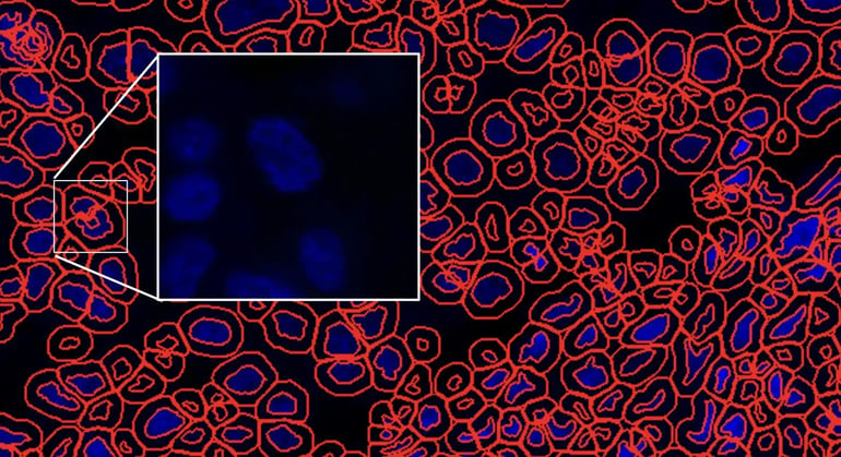 Figure 1. Artifact correction, background, and cell segmentation become more complicated with increasingly higher plex panels, resulting in larger file sizes that require increased compute power.
Mid-plex approaches share many of the same analytical steps, but at a much smaller, more manageable scale—a level where the volume of data and the spatial biology analysis needed to parse it better align with the resources most labs have on hand.
Can you achieve statistical power without breaking the budget?
A well-designed spatial multiplexing experiment is also a resource-efficient one—and the required output can help inform the necessary input.
Generating actionable insights requires statistical significance, correlations, p-values, and the sample sizes that support them. As the cost-per-slide mounts, that means asking an uncomfortable but necessary question early: can you justify significance with five slides' worth of data, or does your study actually need 50? At the cost of a high-plex run per slide—and when those slides represent irreplaceable patient tissue—the math can become prohibitive, fast.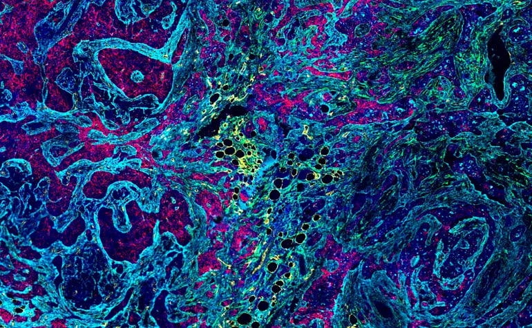
Figure 2. SignalStar® Multiplex IHC panel in infiltrating ductal carcinoma of the breast, illustrating a mid‑plex approach using four markers for tumor, stroma, and microenvironment targets to create a single interpretable dataset. Markers: Lrrc15 (594, green), CD36 (647, yellow), B7H4 ( 750, red), and COL1A1 (647, cyan).
A well-planned mid-plex panel can be a practical way to analyze more slides, generate reproducible data across a larger cohort, and arrive at conclusions you can act on—without spending weeks parsing a massive dataset before you even reach the biology. Whether the end result is a publication, a client deliverable, or simply the next experiment, a focused panel can get you there faster while preserving the quality and interpretability of the underlying data.

Do you need precision and the flexibility to iterate?
There is a significant burden in getting dozens upon dozens of markers to work correctly in tandem. This is a real challenge that the field will hopefully resolve in the years to come, but one that can’t be overlooked today.
Right now, with a more focused marker set, you can optimize a panel to your specific biological questions and tissue conditions and generate signal that is clean and interpretable—and in significantly less time than a high-plex panel would demand. Fewer variables mean faster optimization cycles and a panel that’s inherently more amenable to iteration. With mid-plex, swapping a marker in and out typically doesn’t require rebuilding the experiment from scratch, which can be extremely beneficial, since experiments almost never finish exactly as they were planned!
|
Kelsey Goldman |
“With a more focused marker set, you can optimize a panel to your specific biological questions and tissue conditions and generate signal that is clean and interpretable—and in significantly less time than a high-plex panel would demand.” |

A tighter panel is also easier to standardize and reproduce—across users, instruments, and institutions—which becomes increasingly important as experiments move from a single lab toward broader validation. For translational studies and multi-site collaborations in particular, a mid-plex design can strike a practical balance between biological nuance and operational consistency.
Solutions for Mid-Plex Spatial Biology
If you require something between "a few" and "a great many" markers, CST has a variety of solutions. Selecting the right one depends on your tissue type, imaging platform, and the level of signal amplification your targets require.
SignalStar® Multiplex IHC: Mid-Plex with Built-in Marker Flexibility
SignalStar Multiplex IHC is a customizable, oligo-based assay that amplifies up to 8 markers in formalin-fixed paraffin-embedded tissue (FFPE) tissue, with highly sensitive detection of low-abundance targets and results in 2 days. Antibody panels are designed using the online Panel Builder, delivering validated reproducibility across antibody combinations and the flexibility to swap out targets as experiments evolve. The automated protocol is compatible with leading autostaining platforms, including the BOND RX Automated Stainers by Leica Biosystems and the ONCORE PRO X System by Biocare Medical.
Chimerics: Spatial Multiplexing with Indirect IF & Engineered Antibodies
CST chimerics are recombinant monoclonal antibodies that take the same high-performance rabbit clones you already rely on and re-express them in different host species—horse, feline, chicken, or mouse—enabling up to four-plex IF in frozen and/or FFPE tissue. Each chimeric is validated against its parent rabbit clone to confirm it retains the same specificity and performance, and pairs with standard species-specific, Fc-directed secondary antibodies already common in most labs—no new equipment, buffers, or protocol changes needed.
Fluorophore-Conjugated Primary & Secondary Antibodies
CST offers directly conjugated primary antibodies and secondary antibody-fluorophore conjugates validated for use in FFPE and frozen tissue sections. Primary conjugates enable streamlined, secondary-free detection, while secondary conjugates provide signal amplification and higher signal-to-noise when working with unconjugated primaries. Both are compatible with a broad range of multiplex imaging platforms and suitable for experiments across the plex spectrum.
Carrier-Free Formulations & Custom Labeling Services
For researchers who need antibodies matched to a specific spatial biology platform or labeling chemistry, CST offers BSA- and azide-free carrier-free formulations off-the-shelf, as well as custom conjugation and labeling services. Whether you need a ready-to-conjugate antibody or a fully labeled product built to your requirements, our conjugation team can help you get there without the time and resources required to do it yourself.
Learn More About Multiplexing Solutions from CST
- Blog: Unlock the Missing Marker: Three Ways to Expand Your Multiplex IHC Panel
- Blog: Antibody Compatibility Trifecta: Choosing a Multiplexing Strategy for Reliable Spatial Biology Insights
- Blog: Fluorescent Staining with Multiple Antibodies: Direct vs Indirect Methods for Multiplex Immunofluorescence