



This post is Part 2 of a two-part blog series. Read part 1: What is a TR‑FRET assay and how is it used in drug discovery?
In a previous blog post, we covered the fundamental principles of Time-Resolved Fluorescence (TR) Förster Resonance Energy Transfer (TR-FRET), including how long-lived lanthanide donors like europium enable high-sensitivity, "mix-and-read" assays. Because TR-FRET provides a ratiometric, proximity-based readout in a homogeneous format, it has become a staple in preclinical drug discovery—especially in high-throughput screening (HTS), where automation and data reliability are non-negotiable.
Your specific research focus and biological objectives will dictate your TR-FRET assay design. With a robust toolbox of donor and acceptor conjugates, researchers can tailor TR-FRET assays to the precise demands of any drug discovery program. This versatility allows scientists to configure these assays for diverse therapeutic targets and modalities, generating the data needed to resolve complex research questions across the entire pipeline—from target identification and validation to mechanism of action (MoA) studies.
Here, we explore several types of TR-FRET assays that you can use to ensure your drug discovery program stays on track.
TR‑FRET for Drug Discovery: Protein Quantification, TPD & Competition Assays
By leveraging TR-FRET’s proximity-based readout—where the donor and acceptor must be within 10-100 Å to produce a signal—researchers can design assays for diverse applications. TR-FRET is well-suited for measuring target protein and biomarker levels, protein-protein interactions, receptor-ligand binding, enzyme kinetics, and downstream readouts of small molecule inhibitors (e.g., phosphorylation of downstream target proteins).
-
Advantages: Compared to standard ELISAs, TR-FRET’s main advantage is that it is a no-wash homogenous assay, in which you only need to mix your conjugated antibodies with sample, incubate, and then read. Compared to other no-wash assays such as AlphaLISA, TR-FRET is not affected by ambient light, simplifying handling and minimizing variation found in other platforms. In addition, the TR-FRET signal is long-lasting and can be repeatedly measured, allowing for kinetic analysis. The ratiometric reading of the donor emission relative to the acceptor emission also minimizes well-to-well variation.
-
Considerations: TR-FRET assays are susceptible to the hook effect at high target concentrations, where high levels of the target protein results in a decrease in TR-FRET signal. This can be overcome by diluting the sample into the assay’s linear range. Additionally, while TR-FRET is highly sensitive with a relatively wide dynamic range, other homogeneous assays, such as AlphaLISA, have been reported to be more sensitive with a larger dynamic range.
Here are some of the commonly used assay formats:
Antibody Pair‑Based TR-FRET Assays for Protein Quantification
The closest conceptual cousins to classic sandwich ELISAs, antibody pair-based TR-FRET assays can similarly be used to quantify a target protein or a post-translationally-modified (PTM) protein of interest in samples such as cell or tissue extracts, culture media, and biofluids (e.g., CSF, plasma). These targets, such as disease biomarkers or markers of pathway activation/inhibition, act as functional readouts that can be used across the drug discovery pipeline, from basic research and target identification to high-throughput compound library screens, inhibitor dose-response curves (IC50), lead optimization panels, and mechanism of action studies (MoA).
Unlike traditional ELISAs, TR-FRET assays are performed entirely in solution without wash steps, making them more amenable to high-throughput automation in 384- or 1536-well plates.
Typical design:
-
Donor: Eu³⁺ cryptate conjugated antibody recognizing one epitope on the target.
-
Acceptor: Red‑emitting dye–conjugated antibody recognizing a second epitope.
-
Readout: TR‑FRET signal scales with target concentration in the sample.
When measuring a post-translationally modified (PTM) target, either the donor or acceptor will be a PTM-specific antibody conjugate (e.g., phosphorylation-, ubiquitination-, cleavage-, or acetylation-specific). This includes antibodies that are target- and PTM site-specific (e.g., a conjugate of Phospho-PDGF Receptor beta (Tyr751) (88H8) Mouse Monoclonal Antibody (BSA and Azide Free) #79551), or antibodies that recognize a broad range of post-translationally modified proteins (e.g., Phospho-Tyrosine Mouse Monoclonal Antibody (P-Tyr-100) (trFluor™ Europium Cryptate Conjugate) #36702 ) or Phospho-Tyrosine (D2D1) Rabbit Monoclonal Antibody (trFluor™ Europium Cryptate Conjugate) #28513. An example of this type of assay is shown in Figure 1, where signal increases proportionally with phospho-PDGF Receptor beta (panTyr) abundance across lysate titrations of PDGF-treated NIH/3T3 cells.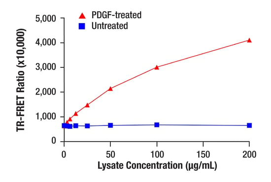
Figure 1. TR-FRET assay was performed using Phospho-Tyrosine Mouse Monoclonal Antibody (P-Tyr-100) (trFluor™ Europium Cryptate Conjugate) and custom conjugate PDGF Receptor β (C82A3) Rabbit Monoclonal Antibody (AlexaFluor® 647 Conjugate) with titrations of PDGF-treated (100 ng/mL, 5 min) NIH/3T3 cell lysate and untreated NIH/3T3 lysate. The relationship between the lysate concentration and the TR-FRET ratio (665 nm/620 nm) is shown. The TR-FRET assay was run in a 384-well white plate and read on a BMG PHERAstar using laser excitation (337 nm) following a 2 hr incubation at room temperature.
The example in Figure 2 shows a TR‑FRET antibody pair assay configured to quantify total protein levels, where signal increases proportionally with target abundance across lysate titrations.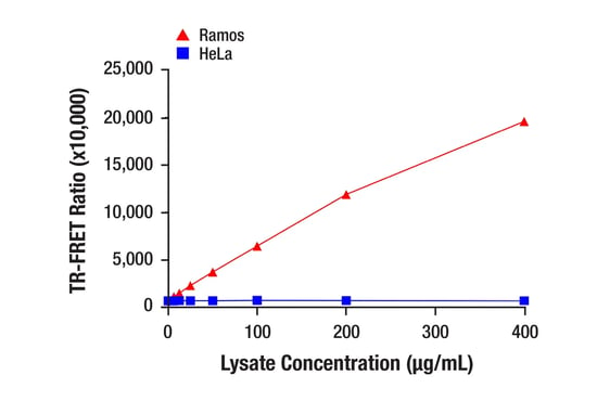
Figure 2. TR-FRET assay was performed using Ikaros (D6N9Y) Rabbit Monoclonal Antibody (trFluor™ Europium Cryptate Conjugate) #76706 and custom conjugate Ikaros (D10E5) Rabbit Monoclonal Antibody (Alexa Fluor® 647 Conjugate) with titrations of Ramos and HeLa cell lysate. The relationship between the lysate concentration and the TR-FRET ratio (665 nm/620 nm) is shown. The TR-FRET assay was run in a 384-well white plate and read on a BMG PHERAstar using laser excitation (337 nm) following a 2 hr incubation at room temperature.
Because these TR-FRET assays mirror ELISA, ELISA-validated antibodies and pairs are the tools that are best suited when starting to build a TR-FRET assay,” explains Jean Loebelenz, Associate Scientist in the Conjugation department at CST. “As with any new assay development, antibody pairs and reagents need to be optimized to ensure performance when transitioning to a different assay platform; however, ELISA-validated antibody pairs provide a solid basis to build TR-FRET assays for a protein or post-translationally-modified protein of interest.”
|
|
Explore Matched Antibody Pairs from CST. Our matched antibody pairs can be custom-conjugated with europium and Alexa Fluor® 647 to fit your specific TR-FRET needs. |
|
TR‑FRET in TPD: Degradation, Ubiquitination, & Engagement Assays
TR‑FRET is particularly powerful in Targeted Protein Degradation (TPD) and other proximity-induced modalities such as PROTACs, molecular glues, LYTACs, AbTACs, RIPTACs, and PhosTACs. TR-FRET assays can be built to screen for degrader molecules and report on a number of readouts relevant to its mechanism of action, such as target abundance, target ubiquitination, and proximity‑driven interactions.
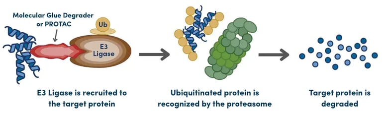 In TPD workflows, the choice of TR-FRET assay depends on which step of the degradation mechanism you need to resolve. Common TPD TR‑FRET assay types include:
-
Degradation Assays: The antibody pair-based TR-FRET format can be used to track loss of target in cells or tissue over time or across degrader dose–response curves. One powerful use case is for high‑throughput ranking of degrader potency and selectivity.
-
Ubiquitination Assays for Mechanism Confirmation: Pairing a target‑specific binder with ubiquitin‑specific antibodies or TUBEs (Tandem-Repeated Ubiquitin-Binding Entities) in a TR‑FRET format can be used to confirm target ubiquitination in response to degrader treatment. Learn more about TUBES-based assays:
-
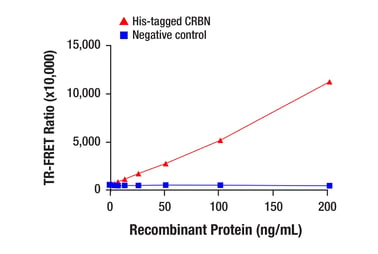
Target Engagement & Ternary Complex Assays: These assays can measure binary binding of degrader to target or E3 ligase or formation of the target–degrader–E3 ligase ternary complex. They often use recombinant proteins with tags (His, FLAG, GST, etc.) along with an anti‑tag TR‑FRET reagent, as shown in Figure 3. This approach allows platform teams to reuse a common set of donor and acceptor‑conjugated partners across multiple targets. If the recombinant proteins do not contain tags or if the assay is measuring endogenous complex formation in cell extracts, antibodies that recognize specific degrader targets and/or E3 ligases (such as CRBN, VHL, c-IAP1) can be used, such as in the example shown in Figure 4.
 |
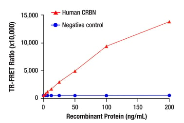 |
| Figure 3. TR-FRET assay was performed using His-Tag (27E8) Mouse Monoclonal Antibody (trFluor™ Europium Cryptate Conjugate) #63381 and custom conjugate CRBN (D8H3S) Rabbit Monoclonal Antibody (Alexa Fluor® 647 Conjugate) with titrations of His-tagged CRBN recombinant protein and a negative control. The relationship between the recombinant protein concentration and the TR-FRET ratio (665 nm/620 nm) is shown. The TR-FRET assay was run in a 384-well white plate and read on a BMG PHERAstar using laser excitation (337 nm) following a 2 hr incubation at room temperature. | Figure 4. TR-FRET assay was performed using CRBN (F4I7F) Rabbit Monoclonal Antibody (trFluor™ Europium Cryptate Conjugate) #69544 and custom conjugate CRBN (D8H3S) Rabbit Monoclonal Antibody (Alexa Fluor® 647 Conjugate) with titrations of human CRBN recombinant protein and a negative control. The relationship between the recombinant protein concentration and the TR-FRET ratio (665 nm/620 nm) is shown. The TR-FRET assay was run in a 384-well white plate and read on a BMG PHERAstar using laser excitation (337 nm) following a 2 hr incubation at room temperature. |
|
|
Explore CST solutions to streamline TPD assay development, including trFluor Europium Cryptate-conjugated antibodies and Alexa Fluor® 647-conjugated antibodies, to support high-throughput screening and MoA studies. |
TR-FRET Competition Assays
TR-FRET can also be configured as a competition or displacement assay, where the signal decreases as an unlabeled molecule competes with or displaces a labeled tracer. This signal drop can be used to quantify analyte abundance, or to measure binding of an unlabeled (unconjugated) compound to a drug target. This format is particularly useful when a traditional antibody pair cannot be used or when assessing small-molecule binding from large compound libraries.
Two scenarios in which competition assays are commonly used:
-
Small Molecule Detection: For targets that are too small to accommodate two antibodies binding to different epitopes, a conjugated tracer (such as a fluorophore‑conjugated analyte) competes with endogenous analyte in the sample for binding to a conjugated antibody. For example, to measure cyclic AMP (cAMP) levels, an assay could use a donor-acceptor pair consisting of a conjugated cAMP antibody and a conjugated cAMP tracer. High TR‑FRET signal is observed when the tracer is bound; increasing concentrations of endogenous cAMP displace the tracer and reduce the signal, enabling dose-dependent, quantitative measurements.
-
Target Engagement/Competitive Binding Screens: In this format, a donor-conjugated antibody (or an anti-epitope tag antibody) binds to the protein of interest while a tracer molecule (a known binder conjugated to an acceptor) also binds to the protein of interest to generate a baseline TR-FRET signal. The addition of unconjugated novel drug compounds that compete for the same binding site displaces the tracer and reduces the signal in a manner that reflects their relative binding affinity. Amenable to high-throughput screening, these types of competition assays typically identify compounds that bind to the same site on the target protein as the tracer molecule. In addition, titration of compound hits identified in this format enables calculation of 𝐼𝐶50 and 𝐾𝑖 values, which can be used to identify compounds with stronger binding affinity.
Setting Up for Success: Where TR‑FRET Fits in Your Workflow
TR‑FRET combines FRET with time‑resolved detection to deliver a homogeneous, no‑wash assay format that is well‑suited for high‑throughput, automated drug discovery workflows. Whether you are quantifying target or biomarker levels, probing PTM state, or dissecting degrader mechanisms of action in TPD, Eu‑based TR‑FRET assays provide a flexible framework that can be adapted to many discovery questions.
Like any immunoassay platform, however, performance ultimately depends on the specificity and affinity of the antibodies you choose, and the robustness of the donor and acceptor conjugations you build on top of them. By starting from well‑validated antibodies and matched antibody pairs, and combining them with optimized Eu‑cryptate donors and red‑shifted acceptors, you can take full advantage of TR‑FRET’s strengths. As you evaluate platforms for your next screening or mechanistic study, TR‑FRET is worth considering wherever you need a sensitive, miniaturizable, and automation‑friendly readout of molecular proximity and protein state.
Learn more about CST Solutions for Sandwich Immunoassay Development