


If you’re running complex multiplex IHC on precious FFPE samples, you already know the tradeoff: every marker you add gets you closer to the biology, but also adds development time, optimization, and risk.
Yet expanding your panel is often the only way to answer the questions that really matter.
-
Are your T cells actually engaging tumor cells or just nearby?
-
Are key checkpoints co-expressed in the same tumor microenvironment or spatially segregated?
-
Is stromal architecture physically or functionally suppressing immune infiltration?
These are spatial biology questions, and they require a flexible, robust antibody panel. The challenge is unlocking that missing marker without burning weeks on assay development and wasting irreplaceable FFPE tissue on trial-and-error.
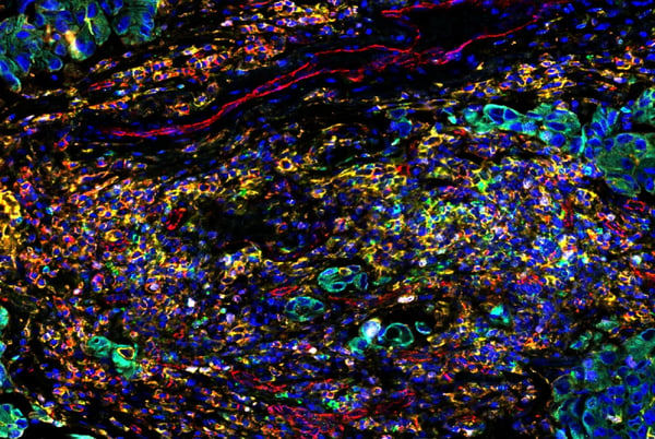
SignalStar oligo-based multiplex immunohistochemical analysis of paraffin-embedded human lung adenocarcinoma using OX40 (ACT35) Mouse Monoclonal Antibody #98785 detected with Anti-mouse IgG SignalStar® Secondary Antibody Kit #50528 (488; green), CD44 (E7K2Y) Rabbit mAb #37259 (594; yellow), CD31 (PECAM-1) (89C2) Mouse mAb #3528 (647; red), Pan-Keratin (C11) Mouse mAb #4545 (750; cyan), Ki-67 (8D5) Mouse mAb #9449 (488; pink), MHC Class II (LGII-612.14) Mouse mAb #68258 (647; orange), and DAPI #4083 (blue). Staining was performed on the BOND RX autostainer by Leica Biosystems.
I started my career working in an IHC core facility, running DAB slides for a wide range of research projects. Although the biological questions varied, one thing was consistent: when timelines were tight and samples limited, we gravitated toward CST antibodies. Not just because of the breadth of the IHC portfolio, but because the antibodies worked.
Today, we’ve translated this broad portfolio into the SignalStar® Multiplex IHC assay, which gives you the reliability of CST reagents in a flexible multiplex workflow that lets you adapt your panel when the biology demands it. SignalStar is an oligo-based multiplex IHC (mIHC) assay validated for the detection of up to eight markers plus DAPI, and delivers quantitative spatial data in about two days.
Here are three practical ways to expand your mIHC panel without starting from scratch.
1. Direct Antibody Conjugates: The Fastest Way to Add Additional Markers
When you need to move quickly, directly conjugated primary antibodies are the simplest way to extend your panel.
This method uses IHC-validated antibodies pre-labeled with fluorophores that you can incorporate into your workflow without changing the core SignalStar chemistry. They’re ready to image right after incubation, and no amplification steps are required.
|
Kelsey Goldman Senior Research Associate |
“If you’re expanding your SignalStar panel using direct conjugates, prioritize the higher abundance targets. Your core SignalStar panel will be amplified, but any additional primaries will not, so you’ll want targets that give you clean signal without amplification.” |

Depending on your panel design, there are two main ways you can use direct conjugates with your SignalStar assay:
-
After SignalStar staining: After completing the final round of SignalStar staining, you can apply the Fluorescence Removal Kit, which removes all fluorescent signal from the staining while leaving the primary antibodies bound. Then, you can add a directly conjugated primary antibody of any channel.
-
During the final staining round: Directly conjugated primaries can also be added during the last round of SignalStar staining, after the ligation step. In this case, you just need to be thoughtful about fluorophore selection and avoid using a direct conjugate with the same fluorophore/channel used in the final SignalStar imaging round (for example, don’t pair an Alexa Fluor® 488 conjugated CD4 antibody with a with a SignalStar Vimentin reagent that is also detected in the 488 nm channel).
The SignalStar assay uses four fluorescent channels—488, 594, 647, and 750 nm—across two iterative rounds of staining. Channels 594 and 647 typically provide the strongest signal and are good choices for lower-abundance antigens, while 488 is more impacted by autofluorescence and 750 generally yields weaker signal. Thinking about fluorophore choice in the context of target abundance helps ensure that any direct conjugates you add will still generate an interpretable signal.
Resource: SignalStar mIHC Antibody to Fluorophore Pairing Considerations
2. SignalStar Secondaries: Fast, Flexible IHC Panel Expansion
If speed is your priority and you need higher sensitivity than direct conjugates, SignalStar secondary antibodies are another powerful route to panel expansion.
The SignalStar workflow uses oligo-tagged primaries and does not rely on secondaries for its core markers. With the SignalStar secondaries, available in both anti‑rabbit and anti‑mouse formats, you can incorporate any rabbit- and/or mouse-derived primary antibody as additional markers in your panel.
 |
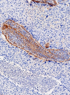 |
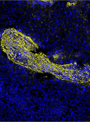
Figure 1. COL1A staining in human squamous cell carcinoma. Right: COL1A detected using the unconjugated primary antibody COL1A1 (E8F4L) Rabbit Monoclonal Antibody #72026 and a traditional DAB IHC protocol. Left: COL1A detected on serial tissue using the same unconjugated primary antibody and the Anti-rabbit IgG & CO-0055-594 SignalStar Secondary Antibody Kit #41483, demonstrating comparable localization with multiplex-compatible fluorescent detection.
Because a large portion of our IHC-validated antibodies are rabbit or mouse recombinant monoclonals, this strategy gives you immediate access to a broad catalog of reagents validated for IHC in FFPE tissues.
Adding a secondary antibody does introduce a few extra steps and, like any panel adjustment, benefits from some optimization. Indirect detection with a labeled secondary also gives you the potential for signal amplification, since multiple secondary antibodies can bind each primary, helping with the detection of lower abundance targets. We recommend titrating your primary antibody and running a serial DAB match to confirm that staining intensity and pattern for the secondary-based marker are consistent with the unconjugated parent antibody.
3. Custom Antibody Conjugation: Add the Exact Marker You Need
Sometimes the biology doesn’t care what’s “pre-validated” and “panel-ready.”
If your study hinges on a specific marker—whether it’s a rare immune subset, a stromal target, or an emerging checkpoint—you can bring it into your SignalStar panel through custom conjugation.
At the core of the SignalStar assay are proprietary oligo tags attached to IHC-validated primary antibodies, enabling amplified fluorescent detection on FFPE tissue. By working with our Antibody Conjugation team, you can convert our IHC-validated primary antibodies into SignalStar-compatible reagents.
Working with our team, you'll select your IHC-validated primary and your desired SignalStar channel. Once those are finalized, we manage the technical specifications for conjugation to specific channels and coordinate the delivery of the DAB serial match.
Custom conjugation is the most flexible option when you need to track a very specific target. Reach out to our custom conjugation experts, and they'll help guide you through the process.
Unlock Spatial Biology Insights — With Room to Expand
Cell Signaling Technology (CST) has one of the broadest portfolios of robustly validated IHC targets in the industry, and SignalStar is built directly on that foundation.
Whether you’re extending your panel with direct conjugates, adding markers through secondary antibodies, or creating a fully custom oligo-conjugated reagent, you’re still working within a framework designed for FFPE tissue and validated by our in-house experts. That means you can expand your panels to follow where the biology leads—new markers, new combinations, new hypotheses—without having to restart validation every time.
In other words, you get the flexibility to iterate quickly and the confidence that your multiplex data will hold up when it matters.
Learn more about SignalStar Multiplex IHC and spatial biology solutions from CST:


