


Pyroptosis is a form of regulated necrotic cell death that ties the danger-sensing capabilities of the innate immune system to gasdermin-mediated membrane permeabilization and inflammatory cell lysis.1 Unlike classic necrotic cell death, which is typically accidental and driven by physical or chemical damage, pyroptosis is a programmed and regulated self-destruct pathway that cells enter in response to damage or stress.2
At the center of pyroptosis is inflammasome signaling—large multiprotein complexes that form in the cytosol and activate inflammatory caspases, cleave gasdermins to initiate pore formation, and trigger the release of pro-inflammatory cytokines.
This blog explores the key mechanisms, pathways, and proteins involved in pyroptosis, along with antibody tools to help distinguish it from other types of cell death and map its role in infection, inflammation, and disease.
|
Explore CST antibody sampler kits for studying pyroptosis, which include sensor, adaptor, caspase, and gasdermin targets to track inflammasome priming, activation, and downstream pyroptotic cell death: |
|
What is Pyroptosis?
Pyroptosis is a type of programmed necrotic cell death that is driven by innate immune sensing pathways that detect pathogen‑associated molecular patterns (PAMPs) or damage‑associated molecular patterns (DAMPs). While early research focused on pyroptosis induced by intracellular infections from bacteria, viruses, fungi, and protozoa, we now know that inflammasome-driven pyroptosis can also respond to “sterile” stimuli such as metabolic stress, crystalline DAMPs (e.g., urate or cholesterol crystals), and protein aggregates to initiate cell death.3
After these dangers are sensed, pyroptosis proceeds through two main pathways:
- Canonical inflammasome‑dependent pathway: In response to stress or damage signals, NLRP3 and other inflammasome sensors assemble with ASC/TMS1 in the cytosol to form inflammasomes and activate caspase‑1, which processes the precursor cytokines pro-L‑1β and pro-IL‑18 and cleaves gasdermin D (GSDMD) to drive pore formation and cell lysis.
- Non-canonical pathway: In response to cytosolic lipopolysaccharide (LPS) from Gram‑negative bacteria, caspase‑4, and caspase‑5 in humans (or caspase‑11 in mice) bind LPS and become activated, then cleave GSDMD to drive pore formation and cell lysis. The non-canonical pathway can also promote canonical NLRP3 inflammasome activation and caspase‑1‑dependent release of inflammatory cytokines.
In both pathways, the result is a rapid loss of membrane integrity, water influx, cell swelling, and lysis, accompanied by a burst of inflammatory cytokines.
Under normal conditions, pyroptosis and inflammasome activation help contain infection, clear damaged cells, and restore tissue homeostasis. When these pathways are repeatedly engaged or fail to resolve, however, inflammasome signaling can contribute to chronic, dysregulated inflammation, persistent IL‑1β/IL‑18 release, and progressive tissue damage. While pyroptosis is most often found in monocytes, macrophages, and dendritic cells—key players in the innate immune system—components of the pathway are found in many tissues. This makes dysregulated inflammasome activity and pyroptosis relevant well beyond acute infection, including in metabolic, cardiovascular, autoimmune, and neurodegenerative diseases.
Moving beyond endpoint cytokine measurements and dissecting the specific danger sensors, activation steps, and executioner mechanisms that define different inflammatory states is a key focus of inflammation and chronic disease research.
Morphological & Molecular Features of Pyroptosis
Cells undergoing pyroptosis show a combination of necrotic and inflammatory characteristics. These include:
- Cell swelling, membrane blebbing, and eventual plasma membrane rupture.
- Formation of gasdermin-mediated membrane pores and release of inflammatory cytokines such as IL‑1β and IL‑18.
- Random DNA fragmentation while the nucleus largely stays intact.
Unlike apoptosis and necroptosis, where chromatin condensation and nuclear fragmentation can be prominently visualized, pyroptotic cells typically lack marked chromatin condensation and instead show limited and scattered DNA breaks.4 Because of this, pyroptotic cells can generate a low, positive signal when analyzed with a TUNEL assay, which may look similar to other lytic or necrotic forms of cell death.
Therefore, additional markers are needed to distinguish pyroptosis from apoptosis and other forms of regulated cell death, such as inflammasome components, inflammatory caspases, gasdermins, and cytokines. In practice, this means combining cleavage‑specific antibodies that distinguish inactive precursors from active signaling and execution events (such as caspase activation, pore formation by gasmerdin D, and mature IL-1β and IL-18) with reagents to visualize inflammasome assembly (such as ASC specks by immunofluorescence). These approaches can be complemented by spatial methods to determine the localization of pyroptotic events within tissues, as well as by flow cytometry to quantify cell death and cytokine production at the single-cell level.The Core Machinery: Inflammasomes & Gasdermins in Pyroptosis
The Core Machinery: Inflammasomes & Gasdermins in Pyroptosis
Pyroptosis is characterized by two protein families: inflammasomes and gasdermins.
The Inflammasome Complex, Activation & Signaling
Inflammasomes are danger‑sensing, multi‑protein machines that link stress and damage signals with inflammatory caspase activation. They typically consist of:
- A cytosolic-pattern recognition receptor (PRR), such as NLRP3 or AIM2-like family members.
- An adaptor protein, ASC/TMS1, which links PRRs to pro‑caspase‑1.
- Pro-caspase‑1, the inflammatory caspase that processes cytokines and gasdermins.
When activated, inflammasomes assemble into large complexes that lead to the formation of visible ASC specks in the cytosol. These specks can be detected by immunofluorescence and are a powerful readout of inflammasome activation. Detecting ASC specks alongside sensor and caspase activation markers can help provide a complete picture of inflammation activation and engagement.
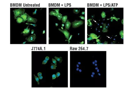
Figure 1. Confocal immunofluorescent analysis of mouse primary bone marrow-derived macrophages (BMDMs) illustrating the translocation of ASC/TMS1 to the inflammasome and ASC speck formation following stimulation with LPS and ATP (white arrows) as a readout of canonical inflammasome activation. BMDMs were either untreated (upper left) or treated with LPS (50 ng/ml, 4 hr, middle) or LPS followed by ATP (5 mM, 45 min, upper right), and J774A.1 (lower left) or Raw 264.7 (lower right) cells, using ASC/TMS1 (D2W8U) Rabbit Monoclonal Antibody (green) #67824. Blue pseudocolor = DRAQ5® #4084 (fluorescent DNA dye).
Gasdermin‑Mediated Pore Formation
Gasdermins are the executioners of pyroptotic cell death. When inactive, gasdermin family proteins are kept in an inhibited state, where their pore‑forming N‑terminal domain is held in check by the C‑terminal domain.
During pyroptosis, inflammatory caspases—caspase‑1, caspase‑4, and caspase‑5 in humans, and caspase‑11 in mice—cleave gasdermin D (GSDMD) to release this N‑terminal fragment, which oligomerizes and inserts into the plasma membrane to form lytic pores. These pores disrupt ion balance, promote water influx, and create channels for the rapid release of cleaved IL‑1β and cleaved IL‑18, linking gasdermin activity directly to both cell lysis and inflammatory signaling.
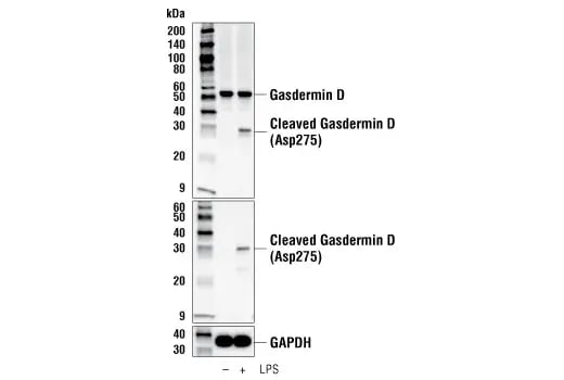
Figure 2. Western blot analysis of THP‑1 cells illustrating gasdermin D cleavage as a readout of pyroptotic execution. Extracts from THP-1 cells were differentiated with TPA (12-O-Tetradecanoylphorbol-13-Acetate) #4174 (50 ng/ml, overnight) and then treated with Lipopolysaccharides (LPS) #14011 (5 μg/ml, 6 hr), using Gasdermin D (E9S1X) Rabbit Monoclonal Antibody #39754 (upper), Cleaved Gasdermin D (Asp275) (E7H9G) Rabbit Monoclonal Antibody #36425 (middle), or GAPDH (D16H11) Rabbit Monoclonal Antibody #5174 (lower).
Different gasdermin family members can be activated by distinct proteases, but GSDMD is the prototypic effector in both the canonical and non-canonical inflammasome‑driven pyroptosis pathways, making antibodies against GSDMD and the inflammatory caspases central tools for detecting and dissecting this pathway in experimental systems. Cleavage‑specific antibodies for caspase‑1, IL‑1β, IL‑18, and gasdermin D are particularly useful here because they can distinguish transcriptional priming and pyroptotic execution in disease‑relevant models across western blotting, immunofluorescence, immunohistochemistry, and flow cytometry. Notably, cleavage‑specific antibodies targeting both the N‑terminal and C‑terminal fragments of gasdermin D provide additional resolution: N‑terminal detection tracks pore‑forming activity, while C‑terminal detection can be useful for identifying gasdermin D released from cells during pyroptosis.
Canonical Inflammasome‑Dependent Pyroptosis
Key Markers: Caspase-1, NLRP3, ASC speckling, IL‑1β, and IL‑18
The canonical inflammasome‑dependent pyroptosis pathway is typically described as a two-step process driven by inflammasome activation of caspase‑1. This pathway is best characterized in innate immune cells such as macrophages and dendritic cells.
- Step 1: Priming & Inflammasome Assembly: Innate immune signals (for example, via Toll-like receptors (TLRs) and NF‑κB) increase the expression of pro-IL‑1β, pro-IL‑18, gasdermin D (GSDMD), and core inflammasome components like NLRP3 and ASC/TMS1. These priming signals can be triggered by infection, but also by chronic inflammatory or metabolic stress.
- Step 2: Caspase‑1 Activation, Cytokines, & Pore Formation: After sensing PAMPs or DAMPs, PRRs such as NLRP3 or AIM2-like family members assemble inflammasomes and bring pro‑caspase‑1 into proximity. Caspase‑1 is activated, then cleaves pro‑IL‑1β and pro‑IL‑18 to create their mature, pro-inflammatory forms, cleaved IL‑1β and cleaved IL‑18, and cleaves GSDMD to release the pore‑forming N‑terminal fragment. GSDMD pores disrupt ion balance, drive water influx, and create channels for cytokine release, leading to cell swelling, membrane rupture, and an intense inflammatory burst.
The signals that drive the initial priming step may be different from the triggers for the second inflammasome activation step, so cells can be transcriptionally primed without fully activating caspase‑1 and initiating pyroptosis.5 Therefore, experimental systems often use separate stimuli to induce transcriptional priming and then trigger full inflammasome assembly and pyroptosis.
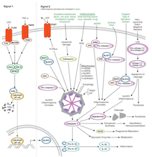 |
Explore the interactive Inflammasome Signaling Pathway diagram, along with associated CST products. |
Non-canonical LPS‑Driven Pyroptosis
Key markers: Caspase‑4, caspase‑5, caspase‑11, cleaved GSDMD, IL‑18, secondary NLRP3/caspase‑1 activation
In the noncanonical pathway, pyroptosis is initiated when cytosolic LPS from Gram‑negative bacteria directly binds and activates caspase‑4 and caspase‑5 in humans, or caspase‑11 in mice. Rather than relying on a PRR–ASC–caspase‑1 inflammasome, these inflammatory caspases act as sensors themselves: LPS binding to their CARD domains drives oligomerization and activation, which then leads to cleavage of gasdermin D (GSDMD). The released GSDMD N‑terminal fragment forms membrane pores, causing the same rapid cell swelling, membrane rupture, and lytic death seen in the canonical pathway.
Noncanonical inflammasome signaling also feeds back into canonical NLRP3/caspase‑1 activation. GSDMD‑mediated pore formation can trigger NLRP3, leading to caspase‑1‑dependent processing and secretion of IL‑1β and IL‑18. As a result, noncanonical pyroptosis is often characterized by both direct GSDMD cleavage by caspase‑4/5/11 and secondary engagement of canonical inflammasome markers.
In experimental systems, combining caspase‑4/5/11 antibodies with GSDMD cleavage assays, as well as cleaved IL‑1β and cleaved IL‑18 readouts, supports activation‑state profiling of LPS‑driven non‑canonical pyroptosis and helps distinguish it from canonical inflammasome‑dependent routes.
Pyroptosis in Disease Research
Pyroptosis and inflammasome signaling are increasingly recognized as central players in infection, chronic inflammatory disease, neurodegeneration, and cancer. Dysregulated NLRP3 activity and repeated pyroptotic cell death can drive persistent IL‑1β/IL‑18 release and tissue damage in conditions such as type 1 diabetes, obesity, cardiovascular disease, and autoimmune disorders, which is why NLRP3 inhibitors are now a major therapeutic focus.
In cancer, pyroptosis is a double‑edged sword. Triggering gasdermin‑mediated lytic death in tumor cells can boost antitumor immunity and help convert “cold” tumors into “hot” ones, but chronic inflammasome signaling and sustained pyroptosis can also support tumor growth and therapy resistance, depending on context.
Neurodegenerative diseases such as Alzheimer’s and Parkinson’s also show evidence of inflammasome activation, ASC speck formation, and pyroptosis in the brain, linking innate immune sensing to neuroinflammation and neuronal loss. Across these areas, reliable assays for NLRP3, ASC, caspase‑1, GSDMD, IL‑1β, and IL‑18—including markers for both the cleaved and un-cleaved forms of the relevant targets—are essential for probing disease mechanisms and testing inflammasome‑targeted therapies.
| Target / Antibody | Application | Reactivity |
| AIM2 (D5X7K) Rabbit Monoclonal Antibody #12948 | WB, IP | H |
| ASC/TMS1 (D2W8U) Rabbit Monoclonal Antibody #67824 | WB, IP, IHC, IF, F | M |
| ASC/TMS1 (E1E3I) Rabbit Monoclonal Antibody #13833 | WB, IP, IHC | H |
| ASC/TMS1 (F8U7P) Rabbit Monoclonal Antibody #78000 | WB, IF, F | H |
| Cleaved Caspase-1 (Asp297) (D57A2) Rabbit Monoclonal Antibody #4199 | WB, IP | M |
| Gasdermin D (E8G3F) Rabbit Monoclonal Antibody #39754 | WB, IP |
H, M, R, Hm |
| Cleaved Gasdermin D (Asp275) (E7H9G) Rabbit Monoclonal Antibody #36425 | WB, IP, IHC | H |
| IL-1 beta (D3U3E) Rabbit Monoclonal Antibody #12703 | WB, IF, F | H |
| Cleaved IL-1 beta (Asp116) (D3A3Z) Rabbit Monoclonal Antibody #83186 | WB, IF | H |
| IL-18 (D4R4T) Rabbit Monoclonal Antibody #54943 | WB | H |
| NALP1 Antibody #56719 | WB | H |
| NLRP3 (D2P5E) Rabbit Monoclonal Antibody #13158 | WB, IP | H |
| NLRP3 (D4D8T) Rabbit Monoclonal Antibody #15101 | WB, IP | H, M |
| NLRP6 (F8J8R) Rabbit Monoclonal Antibody #23077 | WB, IP | M |
| NLRC4 (D5Y8E) Rabbit Monoclonal Antibody #12421 | WB, IP | H |
Additional Resources
Read the additional blog posts in the Mechanisms of Cell Death series:
- Mechanisms of Cell Death: Apoptosis
- Mechanisms of Cell Death: Necrosis & Necroptosis
- Mechanisms of Cell Death: Ferroptosis
Select References
- Galluzzi L, Vitale I, Aaronson SA, et al. Molecular mechanisms of cell death: recommendations of the Nomenclature Committee on Cell Death 2018. Cell Death Differ. 2018;25(3):486-541. doi:10.1038/s41418-017-0012-4
- Escobar ML, Echeverría OM, Vázquez-Nin GH. Necrosis as Programmed Cell Death. In: Cell Death - Autophagy, Apoptosis and Necrosis. InTech.
- Li T, Zheng G, Li B, Tang L. Pyroptosis: A promising therapeutic target for noninfectious diseases. Cell Prolif. 2021;54(11):e13137. doi:10.1111/cpr.13137
- Frank D, Vince JE. Pyroptosis versus necroptosis: similarities, differences, and crosstalk. Cell Death Differ. 2019;26(1):99-114. doi:10.1038/s41418-018-0212-61.
- Cabral JE, Wu A, Zhou H, Pham MA, Lin S, McNulty R. Targeting the NLRP3 inflammasome for inflammatory disease therapy. Trends Pharmacol Sci. 2025;46(6):503-519. doi:10.1016/j.tips.2025.04.007
2626-ICT-93850