Cell cycle regulation is vital to maintain tissue homeostasis and to prevent sustained proliferative signaling and evasion of apoptosis, two of the hallmarks of cancer first described by Doctors Robert Weinberg and Douglas Hanahan. During development, the cell cycle is regulated by three of the four INK4 family members, and their expression subsequently declines with age. In contrast, founding member p16INK4A presents low expression in the early stages of development and steadily increases as we age.1
Given p16’s role in cell cycle regulation and its expression pattern correlating with aging, the protein is a key target for cellular senescence and cancer research.
What role does p16 play in cellular senescence and aging?
Cellular senescence is a response mechanism that prevents cells with extensive DNA damage and harmful genetic mutations from dividing. Thus, cellular senescence is important in preventing cancer and is typically triggered by intrinsic and extrinsic stressors, including telomere shortening, DNA damage, and oxidative stress. Paradoxically, the accumulation of these senescent cells as we age has also been linked to age-related pathologies, including cancer.2
 |
Explore the Senescence Signaling Pathway diagram, along with associated CST products. |
Like the other INK4 family members, p16 is a cell cycle regulator that plays a role in the G1 to S phase transition of the cell cycle. Acting through the retinoblastoma (Rb) pathway, p16INK4A binds to cyclin-dependent kinase 4 and 6 (CDK4 and CDK6) to irreversibly inhibit its activity, prevent transition to S phase, and subsequently cease cellular proliferation. Frequently, p16 is selectively targeted for early inactivation in several cancers, either through point mutations or complete loss, suggesting that its expression is essential for preventing cancer—especially as we age.1,3
This association has been demonstrated in mouse studies, where p16INK4A expression was shown to increase sharply later in life. Whereas the three other members of the INK4 family—p15INK4B, p18INK4C, and p19INK4D—were widely expressed during early development, p16 is suppressed in healthy, young tissue, and expression increases over time.1
The ability of p16 to cease proliferation has been observed as an important role in tumor suppression, and both mutations in and the loss of the gene in its entirety have been linked to several types of cancers. In most cancers, p16 is typically inactivated early on to enable disease progression.2 However, the normal role of p16 remains unclear, and the expression levels are highly variable in several cancer types.3
How does p16INK4A contribute to cancer progression?
Senescent cells can exhibit dramatic changes in their secretome—a process known as senescence-associated secretory phenotype—that include an upregulation and release of several pro-inflammatory cytokines, proteases, and growth factors that can affect the surrounding tissues. As a consequence, this accumulation of senescent cells during aging without their proper removal by the immune system may contribute to the progression of tumor development.
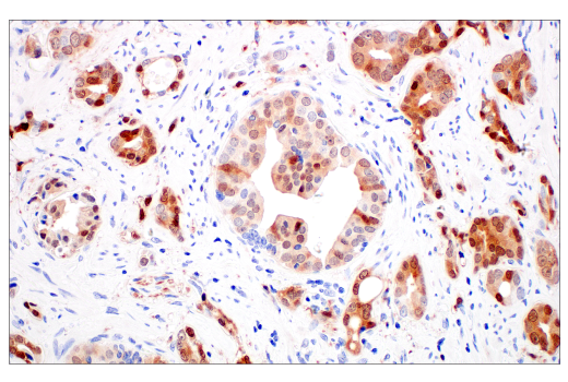
Observations of highly expressed p16 correlate with some types of pRb-negative and malignant cancers, including cervical carcinoma, urothelial carcinoma, glioblastoma multiforme, head and neck squamous cell carcinoma, lung squamous cell carcinoma, prostate adenocarcinoma (Figure 1), and cutaneous melanoma.2,3
 Figure 1. Immunohistochemical analysis of paraffin-embedded human prostate adenocarcinoma using p16 INK4A (BC42) Mouse Monoclonal Antibody #68410.
However, the significance of p16 overexpression remains unclear. Further research is needed to determine whether it is just a futile attempt to prevent cellular proliferation due to a dysfunctional G1-S phase checkpoint, or if this overexpression is eliciting a strong immune-oncogenic response.
An extensive study on p16 expression in 124 different tumor types found a link between HPV infection and strong expression of p16 in certain cancers, including cervical, uterine, and penile.3,5 Even so, this link is inconsistent and additional research is needed to understand the multiple roles that p16 exhibits during tumor progression.
Importance of IHC-Validated Antibodies for Cancer Research
Cancer researchers frequently use immunohistochemistry (IHC) to detect specific biomarkers in a tissue sample for the diagnosis and characterization of cancer. Over 3,000 studies have used IHC to study p16 biomarker expression in normal and tumor tissues. Yet, the role of p16 expression in many cancers remains unclear due to the highly variable results obtained. This variability is likely due to the use of multiple non-validated antibodies, different immunostaining protocols, and non-standardized criteria used to determine p16 expression levels.3
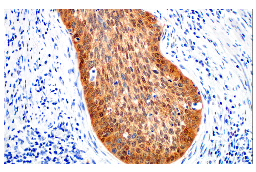
The p16 INK4A (BC42) antibody (Figure 2) from Cell Signaling Technology recognizes endogenous levels of total p16INK4A protein. It has been rigorously validated for IHC using our Hallmarks of Antibody Validation Principles and tested on several commonly studied human tissues, including cervical carcinomas.
 Figure 2. IHC analysis of paraffin-embedded human squamous cell carcinoma of the cervix using p16 INK4A (BC42) Mouse Monoclonal Antibody #68410 performed on the BOND RX Fully Automated Research Stainer by Leica Biosystems.
Since the variation observed in senescence biomarker expression can depend on senescence stimuli, cell type, and timing, it is important to use multiple senescence-associated markers, like p16, for proper identification and characterization.
Given the distinct and variable p16 expression profiles observed in cancer, it is important to have highly validated antibodies for accurate analysis in research studies. The determination of target specificity in IHC analysis requires multiple validation steps. At CST, the steps for validating our IHC antibodies include the use of paraffin-embedded cell pellets (Figure 3) and xenografts of cell lines with known target expression levels, blocking peptides to verify specificity, assessment in relevant mouse models of cancer and human cancer tissue arrays, as well as a variety of other methods.

Figure 3. IHC analysis of paraffin-embedded HeLa cell pellet (left, positive) or MCF7 cell pellet (right, negative) using p16INK4A (BC42) Mouse Monoclonal Antibody #68410, one of the steps in the validation of antibodies for IHC assays at CST.
Rigorous antibody validation and optimization methods that are specific to use in IHC assays provide scientists with confidence in their results while also eliminating the time spent on this process in-house, allowing them to shift focus back to their research.
p16INK4A: A Vital Biomarker for Studying Senescence in Aging & Cancer Research
Due to p16’s expression during cellular senescence, association with aging, and role as a cell cycle regulator, it is a key target for cancer research. To date, there have been a large number of p16INK4A research studies and an observed link between its expression and specific types of malignant cancers, yet the significance and role of p16 expression remains uncertain due to the highly variable results among the studies.
Ongoing research requires the use of IHC-validated antibodies for accurate detection and analysis of its expression to understand the pivotal roles it plays in cellular senescence, aging, and cancer research.
Complete Your Senescence Research Tool Kit
|
Western Blot, IF, IP |
|
|
Senescence Sampler Kit |
|
|
Senescence β-Galactosidase Activity Assay for Flow Cytometry |
Senescence β-Galactosidase Activity Assay Kit (Fluorescence, Flow Cytometry) #35302 |
|
IHC SignalStain Boost |
SignalStain IHC Dual Staining Kit (HRP, Rabbit, Brown / AP, Mouse, Red) #82456 |
Learn More:
- Products and tools to measure cellular senescence
- Learn more about Cell Cycle and DNA Damage and Repair
Select References:
-
Zindy F, Quelle DE, Roussel MF, Sherr CJ. Expression of the p16INK4A tumor suppressor versus other INK4 family members during mouse development and aging. Oncogene. 1997;15:203-211. doi: 10.1038/sj.onc.1201178
-
LaPak KM, Burd CE. The Molecular Balancing Act of p16INK4A in Cancer and Aging. Mol Cancer Res. 2013;12(2):167-83. doi: 10.1158/1541-7786.MCR-13-0350
-
De Wispelaere N, Rico SD, Bauer M, et al. High Prevalence of p16 Staining in Malignant Tumors. PLoS ONE. 2022;17(7):e0262877. doi: 10.1371/journal.pone.0262877
-
Liu Y, Sharpless NE. Tumor Suppressor Mechanisms in Immune Ageing. Curr Opin Immunol. 2009;21(4):431-439. doi: 10.1016/j.coi.2009.05.011
-
Nicolás I, Saco A, Barnadas E, et al. Prognostic Implications of Genotyping and p16 Immunostaining in HPV-positive Tumors of the Uterine Cervix. Mod Pathol. 2020;33:128-137. doi: 10.1038/s41379-019-0360-3
23-CAN-00800


