


Have you ever spent time treating cells, preparing cell extracts, and performing a western blot only to be disappointed when you don’t see the expected signal or change in protein expression? That inevitably leads to the next question—did something go wrong with the experiment? Using control cell extracts as positive and negative controls in your immunoblots can tell you if there is an issue and, if so, help identify the problem.
Did you see no change in protein expression after treating the sample, but you did see a change in signal with the control cell extracts? Then the issue doesn’t lie with your antibody, and you may want to take a closer look at your sample preparation.
Control cell extracts are especially useful if you’re investigating cell death-related events for the following reasons:
- Signal Induction: Cell death pathways are extremely nuanced, making it sometimes difficult to induce the desired cell death pathway in your sample. Different treatments can lead to the activation of different signaling cascades.
- Sample Preparation: It takes experience and the right protocol to know what sample preparation method is optimal.
- Sample Collection: Cell death-related targets often undergo transient post-translational modifications (PTM), like a cleavage event, so you could be preparing extracts at the wrong time point.
Cell Signaling Technology offers a few different control cell extracts, each of which consists of a negative and positive control that can be used for your cell death western blot experiments. The negative control contains either no or lower endogenous levels of the protein or PTM compared to the positive control, which contains higher levels of the induced protein/modification of interest. As a bonus, most of the compounds used by CST to induce cell death in the control cell extracts, like etoposide, chloroquine, and thapsigargin, are also available for you to treat your own cells with if you want to minimize experimental variables as much as possible.
This post explores the best control cell extracts to use as positive or negative controls in your western blots when studying apoptosis, autophagy, and mitophagy.
Which control cell extracts should I use for apoptosis western blots?
Apoptosis is a type of programmed cell death that is mediated through caspase cleavage events as part of the cell death pathway. It is characterized by cell membrane blebbing, cell shrinkage, nuclear fragmentation, chromatin condensation, DNA fragmentation, and mRNA decay. Defects in apoptosis are implicated in cancer, neurodegeneration, cardiovascular disorders, and autoimmune diseases.1 Apoptosis can be chemically induced in cells using either etoposide or cytochrome c.2-3

CST offers two control cell extracts for detecting apoptosis via western blot:
The positive controls for Jurkat Apoptosis Cell Extracts (etoposide) #2043 and Caspase-3 Control Cell Extracts #9663 are produced from total Jurkat cells treated with 25 µM etoposide for 5 hours or the cytoplasmic fraction of Jurkat cells treated with cytochrome c, respectively. Both can be used as control cell extracts for your apoptosis western blots.
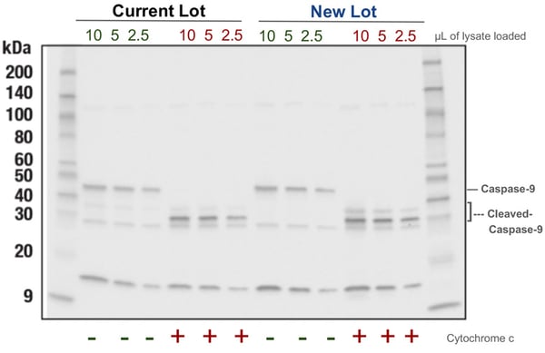
 Figure 1. Representative data from Caspase-3 Control Cell Extracts #9663 new lot testing. Western blot analysis is shown for Caspase-9 Antibody #9502 (top) and Caspase-3 (D3R6Y) Rabbit Monoclonal Antibody #14220 (bottom). As expected, cleavage of caspase-3 and -9 are induced upon cytochrome c treatment (+). Full length forms of both proteins are seen in untreated extracts (-). Lot-to-lot signal is maintained.
Below are some of the targets for which you can use Jurkat Apoptosis Cell Extracts (etoposide) #2403 and the Caspase-Control Extracts #9663 as controls when running a western blot to examine apoptosis.
| Target | Jurkat Apoptosis Cell Extracts (etoposide) #2043 | Caspase-3 Control Cell Extracts #9663 |
| Caspase-2 | ✓ | |
| Caspase-3 | ✓ | ✓ |
| Cleaved Caspase-3 (Asp175) | ✓ | ✓ |
| Caspase-6 | ✓ | |
| Cleaved Caspase-6 (Asp162) | ✓ | ✓ |
| Caspase-7 | ✓ | ✓ |
| Cleaved Caspase-7 (Asp198) | ✓ | ✓ |
| Caspase-8 | ✓ | ✓ |
| Cleaved Caspase-8 (Asp374) | ✓ | ✓ |
| Cleaved Caspase-8 (Asp384) | ✓ | ✓ |
| Cleaved Caspase-8 (Asp391) | ✓ | |
| Caspase-9 | ||
| Cleaved-Caspase-9 (Asp315) | ✓ | ✓ |
| Cleaved Caspase-9 (Asp330) | ✓ | ✓ |
| Cleaved DFF45 (Asp224) | ✓ | |
| Cleaved α-Fodrin (Asp1185) | ✓ | |
| PARP | ✓ | |
| Cleaved PARP (Asp214) | ✓ |
Which control cell extracts should I use for autophagy western blots?
Autophagy is a type of programmed cell death where autophagosomes and lysosomes remove degraded cell components as part of the autophagy signaling pathway. Defects in autophagy have been linked to neurodegenerative diseases and cancer.4 Autophagy can be induced with chloroquine treatment.5

The LC3 Control Cell Extracts #11972 positive control is total cell extracts from HeLa cells treated with 40 µM chloroquine overnight, which induces autophagy. The following are just some of the targets the LC3 Control Cell Extracts #11972 can be used for when studying autophagy.
- LC3A/B
- LC3A
- LC3B
 Figure 2. Representative data of LC3 Control Cell Extracts #11972 new lot testing. Western blot analysis is shown for LC3B (E5Q2K) Mouse Monoclonal Antibody #83506 (left), LC3A (D50G8) Rabbit Monoclonal Antibody #4599 (middle) and LC3A/B Antibody #4108 (right). As expected, LC3 isoforms are induced upon chloroquine treatment (+). Full length forms of LC3 are seen in the untreated extracts (-). Lot-to-lot signal is maintained among the current and new lots of lysates.
Which control cell extracts should I use for mitophagy western blots?
Mitophagy is a type of autophagy specifically used to ensure mitochondrial homeostasis. It occurs when mitochondria become defective due to damage or stress. Defects in mitophagy have been linked to neurodegenerative, cardiovascular, metabolic, and skeletal muscle diseases.6 Mitophagy can be chemically induced with thapsigargin treatment.7
The CHOP Control Cell Extracts #33263 positive control is total cell extracts from C2C12 cells treated with 300 nM thapsigargin for 2 hours and can be used to investigate CHOP expression and other mitophagy targets in your western blot.
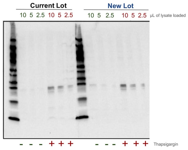
Figure 3. Representative data of CHOP Control Cell Extracts #33263 new lot testing. Western blot analysis is shown for CHOP (L63F7) Mouse Monoclonal Antibody #2895. As expected, CHOP is induced upon thapsigargin treatment (+). At a high exposure, very low signal is seen in the untreated extracts (-). Lot-to-lot signal is maintained among the current and new lots of lysates.
Why should I use control cell extracts from CST?
CST scientists have performed extensive primary and secondary research to identify the most effective methods to chemically induce cell death. From time-course experiments to trying different sample preparation and collection methods, CST control cell extracts are thoughtfully developed and produced to meet your needs. Moreover, the same control cell extracts found in the product catalog are routinely used in-house when validating antibodies.
CST control cell extracts are time-tested, as they have been used internally and externally for years, and are validated with the same stringency applied to all CST products to ensure lot-to-lot reliability. This commitment to product quality makes data interpretation straightforward because you know the target and/or PTM is present.
How are CST control cell extracts validated? First, each batch of cells is tested to ensure there is no mycoplasma contamination. Western blots are then performed in which the new lot of control cell extracts are compared to previous lots for equivalent signal strength and background profiles using multiple different antibodies. If changes in the level of PTMs are expected, then CST standards require testing the new lot of control cell extracts by using, at minimum, one antibody against the total levels of the target protein, and one antibody against the target's PTM, all side-by-side.
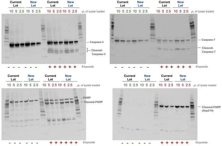
Figure 4. Representative data of Jurkat Apoptosis Cell Extracts (etoposide) #2403 new lot testing. Western blot analysis is shown for Caspase-3 (D3R6Y) Rabbit Monoclonal Antibody #14220 (top left), Caspase-7 Antibody #9492 (top right), PARP Antibody #9542 (bottom left), and Cleaved PARP (Asp214) (D64E10) Rabbit Monoclonal Antibody #5625. Signal is seen as expected in both etoposide treated (+) and untreated (-) extracts. Lot-to-lot signal is maintained across the current and new lots of lysates.
Take Control of Your Cell Death Western Blots
Control cell extracts are tools that prevent you from wasting valuable time trying to determine if your sample preparation or western blot conditions are optimal. CST scientists have worked out the sample treatments, preparation, and collection conditions required to deliver a variety of cell extracts with robust cell death response signals, so you don't have to—giving you the peace of mind that your data interpretation is correct.
Additional Resources
- View the full list of CST control cell extracts.
- Find recommended control treatments by target.
- Explore all the interactive Cell Death Pathways from CST, along with associated CST antibody products.
Select References
- Favaloro B, Allocati N, Graziano V, Di Ilio C, De Laurenzi V. Role of apoptosis in disease.
Aging (Albany NY). 2012;4(5):330-349. doi: 10.18632/aging.100459 - Eischen CM, Kottke TJ, Martin LM, et al. Comparison of apoptosis in wild-type and Fas-resistant cells: chemotherapy-induced apoptosis is not dependent on Fas/Fas ligand interactions. Blood. 1997;90(3):935-943
- Li P, Nijhawan D, Budihardjo I, et. al. Cytochrome c and dATP-dependent formation of Apaf-1/caspase-9 complex initiates an apoptotic protease cascade. Cell. 1997;91(4):479-489. doi:10.1016/s0092-8674(00)80434-1
- Yang Y, Klionksky DJ. Autophagy and disease: unanswered questions. Cell Death Differ. 2020;27(3):858-871. doi:10.1038/s41418-019-0480-9
- Mizushima N, Yoshimori T, Levine B. Methods in mammalian autophagy research. Cell. 2010;140(3):313-326. doi:10.1016/j.cell. 2010.01.028
- Doblado L, Lueck C, Rey C, et al. Mitophagy in Human Diseases. Int J Mol Sci. 2021;22(8):3903. Published 2021 Apr 9. doi:10.3390/ijms22083903
- Wang XZ, Kuroda M, Sok J. et al. Identification of novel stress-induced genes downstream of chop. EMBO J. 1998;17(13):3619-3630. doi.1093/emboj/17.13.3619
%20Formative%20Lab%20Experience/23-fle-89733/whitney-palmer-head-shot.jpeg)


