


When performing spatial analysis of the tumor microenvironment (TME), accurately distinguishing tumor cells from both stromal and immune cells is essential. Correctly identifying epithelial cells is especially critical, as carcinomas, a type of cancer that originates in epithelial tissue, account for 80–90% of all cancer diagnoses1.
Epithelial cancers generally maintain the keratin expression pattern of their cell of origin, even after malignant transformation. As such, researchers often infer tissue origin from keratin immunostaining in primary tumors and metastases, using pan-keratin antibodies to discern tumor cells from the surrounding microenvironment.
But how do you know if a pan-keratin antibody truly recognizes multiple keratin isoforms? Or what if you are struggling due to the mouse-on-mouse background in your pre-clinical model because you’re using a mouse-derived pan-keratin antibody? CST solves both challenges with our new rabbit pan-keratin monoclonal antibody. Rigorously validated for IHC and Western blot, it delivers reliable detection of multiple type I and type II keratins. Plus, because it is rabbit-derived, mouse-on-mouse cross-reactivity is no longer a concern when performing IHC with mouse samples.
<<Jump to the product list at the end of this post>>
What Are Keratins?
Keratins, previously known as cytokeratins2, are the largest group of intermediate filament proteins that serve as key components of the eukaryotic cytoskeleton. Found almost exclusively in epithelial and epithelial-derived cells, keratins maintain cell structure and mechanical stability, protect cells from physical injury, and facilitate intracellular communication with other cytoplasmic components.
Keratins are expressed as obligate heterodimeric pairs, each consisting of a type I (acidic) and a type II (basic) molecule. Because keratin expression patterns vary by cell type, tissue origin, and differentiation state, they are widely used as markers of epithelial identity and cell lineage. Such nuanced expression is especially useful for studying carcinomas, which typically maintain the same keratin expression pattern as their cell of origin (Figure 1).
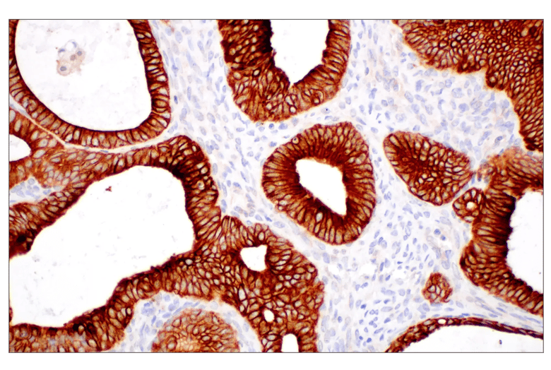
Figure 1. Immunohistochemical analysis of paraffin-embedded human endometrioid adenocarcinoma using Pan-Keratin MultiMab® Rabbit Monoclonal Antibody mix #96963.
Is Your Keratin Coverage Pan-ning Out?
To ensure reliable results when using pan-keratin as a marker of epithelial-derived tumor cells, it is critical to confirm that your pan-keratin antibody is providing the comprehensive coverage you need. This requires application-specific testing and the use of more than one type of validation assay. Just because a variety of keratins are recognized via western blot does not guarantee the same via IHC.
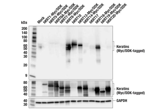
In the example below, Pan-Keratin (C11) Mouse Monoclonal Antibody #4545, an established and well-used clone, recognizes a number of keratins using western blot, but only Keratin 10 with IHC (Figure 2).
_Blog_fig1_web.jpg?width=780&height=459&name=_4545_ihc_Keratin(C11)_Blog_fig1_web.jpg)
Figure 2. Validation of Pan-Keratin (C11) Mouse Monoclonal Antibody #4545. This established clone is proven to recognize multiple keratins via western blot (top). However, it only detects keratin 10 when used for IHC (bottom), highlighting that western blot validation alone is not sufficient to ensure IHC performance.
The Pan-Keratin MultiMab® Rabbit Monoclonal Antibody mix #96963 is validated to detect multiple type I and type II keratins by both western blot and IHC (Figure 3), giving you confidence that it recognizes epithelial-derived tumor cells from multiple cell lineages in both applications.
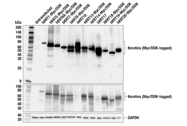
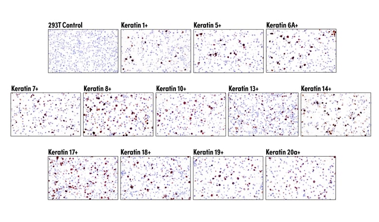
Figure 3: Validation of the Pan-Keratin MultiMab® Rabbit Monoclonal Antibody mix #96963. The clone recognizes multiple keratins via western blot (top) and IHC (bottom), confirming performance for both applications.
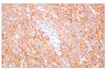
Additionally, the Pan-Keratin MultiMab Rabbit Monoclonal Antibody mix is rabbit-derived, eliminating the need for mouse-on-mouse blocking steps in your IHC protocol and concerns that blocking may not fully prevent all mouse-on-mouse interactions. The result? Clean pan-keratin data on your mouse samples with a protocol that doesn’t require a blocking step (Figure 4).
 |
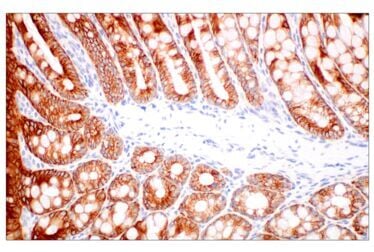 |
Figure 4. Immunohistochemical analysis of paraffin-embedded Renca syngeneic tumor (left) and paraffin-embedded mouse colon (right) using Pan-Keratin MultiMab® Rabbit Monoclonal Antibody mix #96963. Mouse-on-mouse background is eliminated using a rabbit-derived pan-keratin antibody.
And to facilitate your multiplex IHC and spatial biology studies, the Pan-Keratin MultiMab® Rabbit Monoclonal Antibody mix is also available conjugated to Alexa Fluor® 488, Alexa Fluor® 555, and Alexa Fluor® 647, as well as a chimeric antibody re-engineered with a feline backbone for detection with an alternate secondary.
Avoid Analysis Blind Spots: Pan-Keratin vs Single-Isoform Detection
While single isoform antibodies, such as those targeting cytokeratin 7 or cytokeratin 19, can effectively detect epithelial cells in specific tissues like gastrointestinal and ductal tracts, they cannot detect all epithelial or epithelial-derived tumors because no single keratin is universally expressed. Poorly differentiated tumors and those undergoing epithelial-mesenchymal transition (EMT) frequently downregulate keratins, rendering 1- or 2-keratin panels insufficient. Additionally, some cancers, including renal cell and hepatocellular carcinomas may be pan-keratin negative; in such instances, including a secondary marker like CD44 or EpCAM can be helpful.
The Pan-Keratin MultiMab® Rabbit Monoclonal Antibody mix #96963 provides broad coverage of multiple type I and type II isoforms, increasing the likelihood of detecting all carcinoma cells in a sample, including irreplaceable cancer research tissues.
Additional advantages of pan-keratin detection include the ability to:
- Conserve precious panel space in multiplex IHC/IF by reliably marking the entire epithelial compartment with a single channel.
- Serve as a tissue architecture anchor, enabling accurate spatial interpretation of co-stained markers (e.g., immune checkpoints, proliferation markers, and stromal proteins).
- Detect rare epithelial cells in non-epithelial contexts (e.g., micro-metastatic cells in lymph nodes), where surrounding cells lack any keratin expression.
Spatial Analysis of Carcinomas by mIHC: The Multi-Faceted Role of Pan-Keratin
Because pan-keratin antibodies enable researchers to reliably distinguish epithelial cells from other cells in the surrounding milieu, they have utility for many different research applications. These include the following:
Defining the Tumor Compartment and Mapping the Tumor Microenvironment
Keratins are expressed exclusively in epithelial and epithelial-derived cells—not in immune cells, fibroblasts, or endothelium. As such, pan-keratin staining immediately separates tumor parenchyma from stroma in a mixed tissue section, creating the foundational cell classification layer for any multiplex TME panel. Additionally, pan-keratin staining allows for quantification of immune infiltration within vs. outside tumor islands, as well as supports analysis of immune exclusion, spatial proximity, and tumor-immune interactions.
Use pan-keratin staining to distinguish tumor cells from many other cell types (Figures 5 and 6), including:
- Stroma structural cells: CAFs, myofibroblasts, pericytes
- Stroma support cells: cancer-associated adipocytes, mesenchymal stem/stromal cells, mast cells
- Immune cells: TILs, B cells, NKs, macrophages, dendritic cells, MDSCs, neutrophils
- Vascular lymphatic cells: tumor endothelial cells, lymphatic endothelial cells
Multiplex Tumor Compartment Staining with Alexa Fluor®-Conjugated Primary Antibodies.
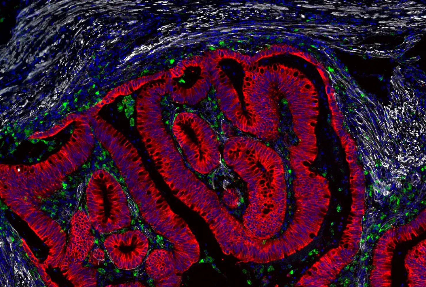
Figure 5. Immunohistochemical analysis of paraffin-embedded human colon adenocarcinoma using Pan-Keratin MultiMab® Rabbit Monoclonal Antibody mix (Alexa Fluor® 647 Conjugate) #34207 (red), CD68 (D4B9C) Rabbit Monoclonal Antibody (Alexa Fluor® 488 Conjugate) #24850 (green), alpha-Smooth Muscle Actin (D4K9N) Rabbit Monoclonal Antibody (Alexa Fluor® 555 Conjugate) #60839 (white), and DAPI #4083 (blue). CD68 identifies tumor-associated macrophages.
Tumor Compartment Staining with Chimeric Primary Antibodies and Alexa Fluor®-Conjugated Secondary Antibodies.
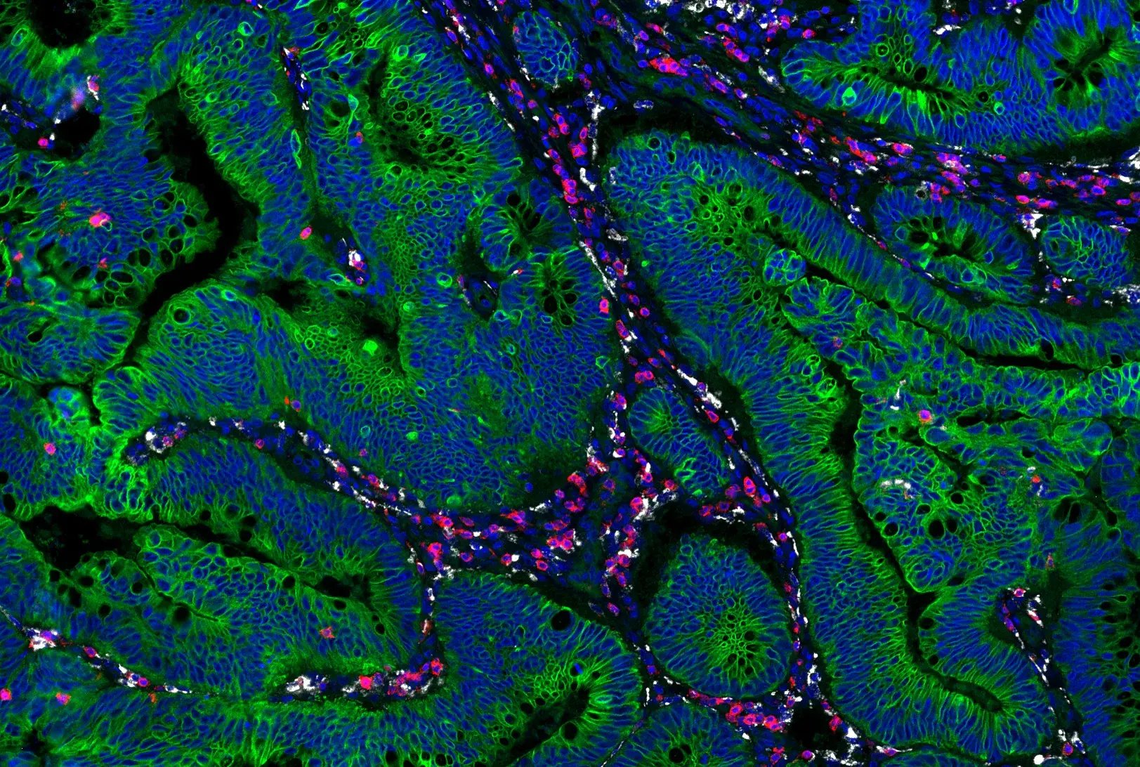
Figure 6. Immunohistochemical analysis of paraffin-embedded human colon adenocarcinoma using Pan-Keratin MultiMab® Feline Chimeric Monoclonal Antibody mix #52203 (green), CD3 epsilon (D7A6E) Mouse Chimeric Monoclonal Antibody #30099 (red), CD68 (D4B9C) Rabbit Monoclonal Antibody #76437 (white), and DAPI #4083 (blue). Rabbit primary antibody is detected using an anti-rabbit Fc-specific secondary antibody CD68 identifies tumor-associated macrophages while CD3 identifies T cells.
EMT Studies
A hallmark of EMT in cancer progression is the downregulation of keratin as keratin
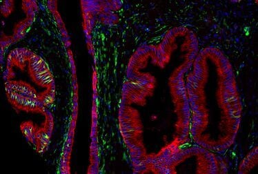
filaments are replaced by vimentin filaments to facilitate metastasis. By co-staining pan-keratin with vimentin and E-cadherin, researchers can classify cells along the epithelial-mesenchymal spectrum within the same section and can spatially map hybrid E/M phenotype cells relative to the invasive front (Figure 7).
Multiplex EMT Staining with Chimeric Primary Antibodies and Alexa Fluor®-Conjugated Secondary Antibodies.
 |
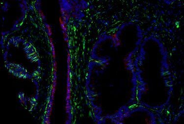 |
Figure 7. Immunohistochemical analysis of paraffin-embedded human prostate adenocarcinoma using Pan-Keratin MultiMab® Feline Chimeric Monoclonal Antibody mix #52203 (left, red), E-Cadherin (D21H3) Rabbit Monoclonal Antibody #3195 (right, red), with Vimentin (D21H3) Mouse Chimeric Monoclonal
Antibody #29066 (green), and DAPI #4083 (blue). Rabbit primary antibody is detected using an anti-rabbit Fc-specific secondary antibody. Lost or downregulated E-cadherin is a sign cells are losing their stationary, structured, and differentiated state, and vimentin identifies mesenchymal cells or cells that have transitioned to a mesenchymal state.
CST Pan-Keratin Portfolio
At CST, we validate all of our antibodies in-house using rigorous, application-specific testing—and the Pan-Keratin MultiMab® Rabbit Monoclonal Antibody mix #96963 is no exception. Proven to detect multiple type I and type II keratins via IHC, the pan-keratin antibody ensures you can confidently discern tumor cells in the TME. Furthermore, if you are working with pre-clinical mouse models, the Pan-Keratin MultiMab Rabbit Monoclonal Antibody mix eliminates the need for mouse-on-mouse blocking, meaning shorter workflows and faster time to results.
To maximize flexibility in your multiplex IHC/IF panel design, explore Pan-Keratin MultiMab Rabbit Monoclonal Antibodies, available as direct Alexa Fluor® conjugates and chimeric formats, along with corresponding secondary antibody offerings.
- Pan-Keratin MultiMab® Rabbit Monoclonal Antibody mix (Alexa Fluor® 488 Conjugate) #57336
- Pan-Keratin MultiMab® Rabbit Monoclonal Antibody mix (Alexa Fluor® 555 Conjugate) #78162
- Pan-Keratin MultiMab® Rabbit Monoclonal Antibody mix (Alexa Fluor® 647 Conjugate) #34207
- Pan-Keratin MultiMab® Feline Chimeric Monoclonal Antibody mix #52203
- Goat Anti-Feline (IgG) (H+L) Antibody (Alexa Fluor® 647 Conjugate) #76333
- Goat Anti-Feline IgG, Fc Fragment Antibody (Texas Red Conjugate) #99224
- Goat Anti-Feline IgG, Fc Fragment Antibody (FITC Conjugate) #39075
Additional Resources
- Resource Center: Multiplex Solutions for Spatial Biology
- Blog: Anything but Rabbit: Why Multiplex with Chimeric Antibodies?
- Blog: Antibody Compatibility Trifecta: Choosing a Multiplexing Strategy for Reliable Spatial Biology Insights
- Blog: Key Epithelial-Mesenchymal Transition (EMT) Markers in Cancer
References
- U.S. National Institutes of Health, National Cancer Institute. Seer Training Modules: Cancer Classification. Accessed September 10, 2026. https://training.seer.cancer.gov/disease/categories/classification.html.
- Schweizer J, Bowden PE, Coulombe PA, et al. New consensus nomenclature for mammalian keratins. J Cell Biol. 2006;174(2):169-174. DOI: 10.1083/jcb.200603161.
26-HMC-18050