


Non-communicable diseases such as neurodegeneration, metabolic disorders, and autoimmune disease are growing in incidence, highlighting the urgent need to develop new treatments and therapies. Unlike pathogen-driven diseases, which can be tackled by eliminating the infectious agent or enhancing patient immunity, non-communicable diseases often require targeting of multiple dysregulated endogenous molecules that drive chronic sterile inflammation. The inflammasome is a central player in the inflammatory process, serving to trigger an appropriate response to danger signals and maintain health. However, the inflammasome has a split personality and can also exacerbate disease pathogenesis if it is not regulated correctly. CST offers an extensive portfolio of antibodies validated in different assays to enable study of the inflammasome in different contexts.
Canonical Inflammasome Activation
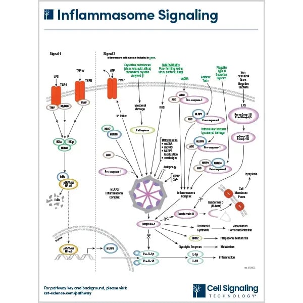
The best-characterized inflammasome is the NLRP3 complex, which consists of the NLRP3 PRR, the ASC adaptor protein, pro-caspase-1, and the serine-threonine kinase NEK7. It is activated via a two-step process, known as the canonical pathway. First, PAMP- or DAMP-mediated activation of TLR4 or TNFR induces NF-κB signaling, leading to increased expression of NLRP3 and the pro-inflammatory cytokine precursors pro-IL-1β, and pro-IL-18; this step is known as priming. Next, NLRP3 associates with ASC and pro-caspase-1, triggering auto-cleavage into active caspase-1 and driving processing of IL-1β, IL-18, and the pore-forming executor of pyroptosis, Gasdermin D (GSDMD); this step is referred to as activation.
Explore our NLRP3 antibodies and antibodies targeting other important PRRs, including NLRP6, NLRC4, AIM2, and Pyrin.
Relevant Pathway
|
Inflammasome SignalingInflammasomes are complex, multi-protein structures that assemble in the cytosol of both specialized immune cells (e.g., macrophages and monocytes) and non-immune cells (e.g., epithelial cells and keratinocytes) following exposure to a danger signal, such as a pathogen- or damage-associated molecular pattern (PAMP/DAMP). They typically consist of a pattern recognition receptor (PRR), a nucleotide-binding domain and leucine-rich-repeat (NLR) or AIM2-like receptor (ALR) family member, an adaptor protein (ASC), and pro-caspase-1, with different inflammasome complexes having different PRRs and activation triggers. |
Non-Canonical Inflammasome Activation
In addition to the classic canonical pathway, a small subset of inflammasome sensors—namely human caspase-4, human caspase-5, and murine caspase-11—can act as both PRRs and effectors to form a non-canonical inflammasome. Specifically, instead of associating with the ASC adaptor, these caspases directly bind lipopolysaccharide (LPS) via their caspase recruitment domains (CARDs) to mediate their own oligomerization and autoproteolytic cleavage.
The Inflammasome and Disease
Inflammasome dysregulation has been implicated in many different diseases, including neurodegeneration, metabolic disorders, and autoimmunity. To better understand how the inflammasome might be targeted to treat these conditions, researchers are investigating inflammasome signaling pathways with methods that allow for quantifying protein cleavage, measuring cytokine release, and monitoring spatial localization.
Keeping the Brain on Fire: Misfolded Proteins and Microglial NLRP3 Activation
Chronic inflammasome activation, mainly via the NLRP3 axis, is known to drive chronic neuroinflammation, leading to synaptic loss and neuronal death. This phenomenon has been widely observed in Alzheimer’s disease (AD), where extracellular Amyloid-β (Aβ) fibrils act as DAMPs and are phagocytosed by microglia to cause lysosomal rupture and NLRP3 assembly. Interestingly, NLRP3 inflammasome deficiencies may play a protective role in AD by skewing microglial cells toward an M2 (anti-inflammatory) phenotype that correlates with decreased Aβ deposition1.
While the inflammasome impact on Parkinson’s Disease (PD) is less well-established, researchers have discovered that intracellular aggregates of α-synuclein and mutant Huntingtin proteins trigger mitochondrial stress and reactive oxygen species (ROS) that directly drive the neuroinflammatory cascade. Additionally, elevated levels of IL-1β and IL-18 resulting from activation of caspase-1—a key mediator of neuroinflammation in the PD brain—have been detected in PD models and patient samples, including cerebrospinal fluid (CSF)2.
-
Learn more about CST Products: NLRP3, Pro-Caspase-1, Cleaved Caspase-1, ASC
○ (IF, IHC & WB): NLRP3, ASC, IL-18, and Cleaved Caspase-1
○ (ELISA): IL-1b and IL-18 can be used to detect the activation of the inflammasome.
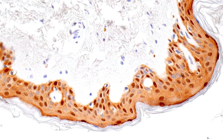 Figure 1. Immunohistochemical analysis of paraffin-embedded normal human skin using IL-18 (E3G8R) Rabbit mAb.
Metabolic Stress as a Danger Signal
Endogenous metabolites can overload cellular homeostasis and cause chronic sterile inflammation in various metabolic diseases. For example, in diabetes, high glucose levels drive inflammation in tissues including the pancreatic islets and kidneys. Notably, individuals with type I diabetes have been found to exhibit higher serum IL-1β and IL-18 levels compared to healthy subjects, potentially due to dysregulation of the NLRP3 inflammasome3. However, to date, the impact of the inflammasome on diabetes has mostly been investigated in mouse models.
Besides NLRP3, other PRRs thought to be involved in metabolic diseases include NRLP1, NLRP6, NLRC5, NLRP12, and NLRP2.
-
Learn more about CST Products: NLRP3, NLRP6, Phospho-NF-κB p65, Total NLRP3, Cleaved Gasdermin D, and IL-1β.
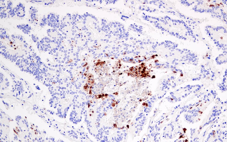
Figure 2. Immunohistochemical analysis of paraffin-embedded human colon carcinoma using Cleaved Gasdermin D (Asp275) (E7H9G) Rabbit mAb in the presence of non-cleaved Gasdermin D peptide.
When the Brake Fails: Inappropriate Responses to Metabolic Stress and Autoantibody-Driven Inflammasome Activation
Inflammasome dysregulation has been linked to several autoimmune diseases, including rheumatoid arthritis (RA). In RA patients, elevated serum urate levels (hyperuricemia) are associated with the formation and accumulation of monosodium urate (MSU) crystals in articular and periarticular tissues. Here, the MSU crystals serve as DAMPs, triggering the NLRP3-driven release of pro-inflammatory cytokines that amplify local inflammation4.
Inflammasome dysregulation is also implicated in Systemic Lupus Erythematosus (SLE), a condition characterized by the production of numerous autoantibodies. These include circulating anti-dsDNA autoantibodies with the ability to form immune complexes that enable them to enter cells and drive mitochondrial ROS production, promoting systemic inflammasome assembly5.
Learn more about CST Products: Spatial immune markers (CD14, CD3), Total/Cleaved Caspase-1, ASC, IL-1b 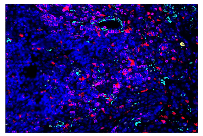
Figure 3. SignalStar® oligo-based multiplex immunohistochemical analysis of paraffin-embedded human melanoma using ASC/TMS1 (E1E3I) Rabbit Monoclonal Antibody (594; yellow), alpha-Smooth Muscle Actin (D4K9N) Rabbit Monoclonal Antibody (488; cyan), Ki-67 (8D5) Mouse Monoclonal Antibody (594; yellow), CD3 epsilon (D7A6E) Rabbit Monoclonal Antibody (647; magenta), and DAPI #4083 (blue). Staining was performed on the BOND RX autostainer by Leica Biosystems.
Conclusion
Whether you are studying the microglial response to Amyloid-β, kidney function in hyperglycemia, or autoantibody-mediated joint destruction, the inflammasome pathway remains a key area of research. To move your studies forward with confidence, choosing application-specific antibodies and a vendor with a comprehensive portfolio of products is essential. At CST, we offer an extensive selection of antibodies for inflammasome research, all of which are rigorously validated according to the CST Hallmarks of Antibody Validation.
Select References
-
Heneka MT, Kummer MP, Stutz A, et al. NLRP3 is activated in Alzheimer’s disease and contributes to pathology in APP/PS1 mice. Nature.2013;493(7434):674-678.
-
Nguyen LTN, Nguyen HD, Kim YJ, et al. Role of NLRP3 Inflammasome in Parkinson's Disease and Therapeutic Considerations. J Parkinsons Dis. 2022;12(7):2117-2133.
-
Wang J, Shen X, Liu J, et al. High glucose mediates NLRP3 inflammasome activation via upregulation of ELF3 expression. Cell Death Dis. 2020;11,383.
-
Du L, Zong Y, Li H, et al. Hyperuricemia and its related diseases: mechanisms and advances in therapy. Signal Transduct Target Ther. 2024;9(1):212.
-
Liu Y, Tao X, Tao J. Strategies of Targeting Inflammasome in the Treatment of Systemic Lupus Erythematosus. Front Immunol. 2022;13:894847.
26-ICT-51550
26-bre-69300
fff26-HMC-18050