


Proteins to Watch in Huntington's Disease Research
When developing assays to study Huntington's disease (HD), scientists have a rare advantage: they know the exact genetic cause. But that hasn't made the biological mechanism any easier to tackle. Despite decades of research, effective treatments remain limited for this progressive degenerative disease.2 HD is inherited in an autosomal-dominant manner, passing on a pathogenic CAG repeat within exon 1 of the HTT gene, which results in production of the mutant huntingtin protein (mHTT) with an expanded polyglutamine tract.1 Over time, this mutation drives protein misfolding, aggregation, and neuronal dysfunction, and the brain’s striatum bears the brunt of the damage as medium spiny neurons (MSNs) are progressively lost.1,2
Because HD follows autosomal dominant inheritance, the mutation is detectable decades before symptoms appear, giving clinical researchers a rare opportunity to study neurodegeneration at its earliest, most treatable stages.2 This head start means scientists can develop assays to examine relevant biomarkers in a genetically defined population and assess the impact of emerging therapeutics, like treatments that lower mHTT.2
Making the most of that opportunity hinges on choosing the right antibody biomarkers for your preclinical compound screens. Below, we've outlined key molecular targets in HD research—from the huntingtin protein itself to downstream markers of striatal dysfunction, neuronal injury, and metabolic failure—making it easier to select the right panels of validated Cell Signaling Technology (CST) antibodies for the most informative preclinical readouts.
How Huntingtin Disrupts Protein Interactions
Protein accumulation is the defining molecular feature of HD. As mHTT misfolds and aggregates, it disrupts proteins that normally regulate neuronal signaling, intracellular trafficking, and synaptic organization.1,2 Antibodies that target huntingtin and its interacting partners are essential tools for studying the pathobiology of HD—and for evaluating the growing number of HTT-lowering therapeutic strategies in development.

#1. Huntingtin (HTT / mHTT)
Huntingtin is a large (~350 kDa) cytosolic protein expressed throughout the body, with the highest expression in the brain—particularly in the striatum, neocortex, hippocampal formation, and cerebellar cortex.1In HD, the expanded polyglutamine tract in mHTT promotes misfolding and proteolytic cleavage, leading to aggregation-prone N-terminal mHTT fragments, which subsequently impact transcription, axonal transport, mitochondrial homeostasis, and autophagy.1,2
As HTT-lowering strategies continue to be investigated, measuring mHTT in CSF and plasma has become a key pharmacodynamic endpoint—one that requires antibodies with proven sensitivity and specificity for accurate readouts in bead-based immunoassays and ELISAs.2 A 2024 consensus platform study comparing 20 commercial huntingtin antibodies found significant variability in performance across reagents, underscoring the importance of using rigorously validated tools for any assay-based mHTT detection workflow.3 Antibodies developed at CST undergo unparalleled, application-specific validation using a multilayered strategy before they reach the market—so researchers can trust that our antibody tools will consistently deliver specific, reproducible results in their recommended applications.
CST offers a portfolio of highly validated recombinant monoclonal antibodies that target distinct epitopes across the full-length huntingtin protein, from the N-terminal region near the polyQ domain through the N-HEAT repeats, bridge domain, and C-HEAT repeats. They enable detection across a range of disease models—including human or mouse tissues and primary cells, such as iPSC-derived neurons—and research applications, including ELISAs, high-content screening (HCS), and immunofluorescence (IF).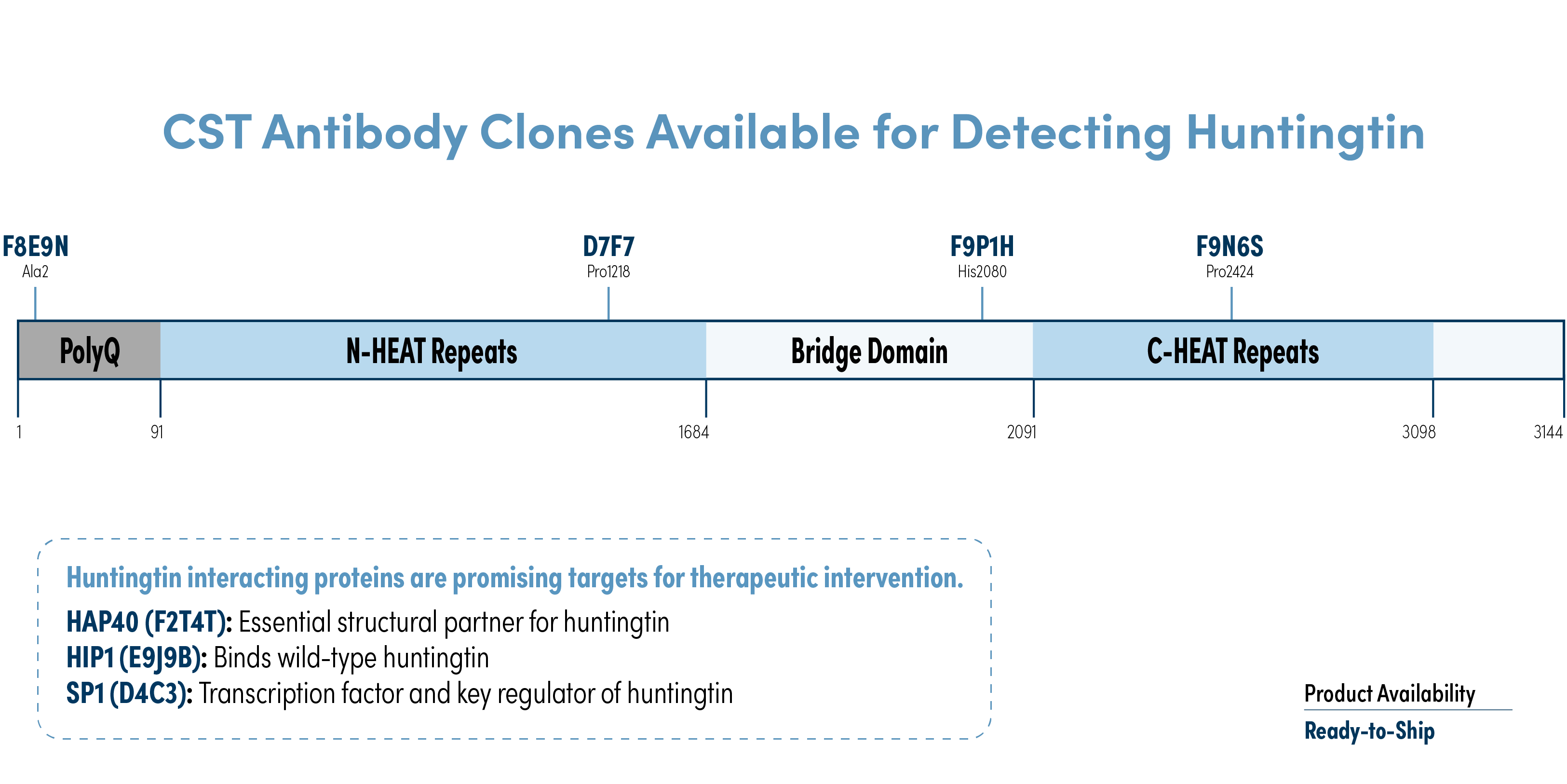
- Huntingtin (D7F7) Rabbit Monoclonal Antibody (Biotinylated) #93535 – ELISA+; H, M, R
- Huntingtin (D7F7) Rabbit mAb (BSA and Azide Free) #31873 — WB, IF; H, M, R
- Huntingtin (F8E9N) Rabbit mAb #73232 — WB, IHC, IF; H, M, R
- Huntingtin (F9P1H) Mouse mAb #58445 — WB, IF; H, M, R
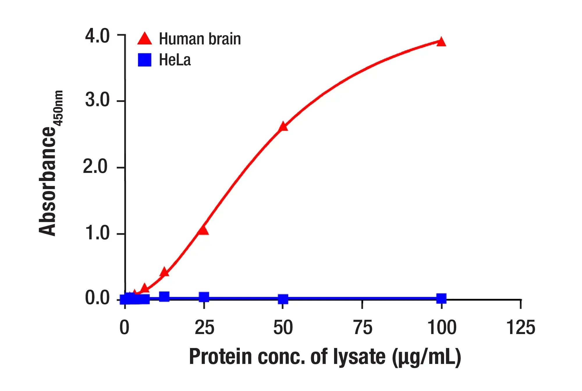
Figure 2. An ELISA was performed on a plate coated with a synthetic peptide of the human huntingtin protein corresponding to residues around Pro1218. The figure shows the relationship between the concentration of biotinylated (blue) or non-biotinylated (red) Huntingtin (D7F7) Rabbit mAb and the absorbance at 450 nm on a peptide-coated plate. Streptavidin-HRP #3999, TMB substrate #7004, and STOP Solution #7002 were used for color development.
#2. HAP40
HAP40 is an essential structural partner of HTT, required for HTT stability and proper intracellular function, and its levels decline alongside HTT during HD progression.
- HAP40 (F2T4T) Rabbit mAb #53720 — WB; H, M
#3. HIP1
HIP1 links huntingtin mechanisms to endocytic trafficking and cell survival signaling. In HD, mHTT shows reduced affinity for HIP1, potentially freeing it to engage with pro-apoptotic pathways linked to striatal neuronal vulnerability.
- HIP1 (E9J9B) Rabbit mAb #90830 — WB; H, M, R
#4. PSD-95
PSD-95 is a postsynaptic scaffolding protein that’s essential for organizing glutamatergic receptor complexes at synapses. In HD models, huntingtin directly influences PSD-95 clustering in striatal neurons, and progressive loss of synaptic proteins, including PSD-95, has been documented as the disease advances.⁴
- PSD95 (D27E11) Rabbit mAb #3450, Mouse Chimeric mAb #31225, Horse Chimeric mAb #19622, or Feline Chimeric mAb #90708 — IF; H, M, R
- PSD95 (7E3) Mouse Monoclonal Antibody (BSA and Azide Free) #37350 — WB, IF; H, M, R
- Phospho-PSD95 (Ser295) (A8F8Z) Rabbit Monoclonal Antibody #45737 — WB; H, M, R
 Many CST biomarker antibodies are now available in even more host species—allowing you to build panels for multiplexing and high-content screening with ease. |
Tracking Neuronal Loss: NfL and NeuN in HD Research
As HD progresses, mHTT toxicity and cellular dysfunction become apparent and observable as neuronal injury and loss. The biomarkers below can help to examine and quantify neurodegeneration across tissue and biofluid samples, and are well established in longitudinal HD research.5, 9
#5. Neurofilament Light Chain (NfL)
NfL is currently the most clinically well-supported fluid biomarker for HD.5 Plasma NfL rises in premanifest gene carriers up to a decade before motor symptom onset, and a 2025 systematic review with meta-analysis concluded that NfL meets established evidentiary guidelines as a prognostic biomarker in premanifest HD—a distinction no other fluid biomarker has yet achieved.5 NfL serves as a clear benchmark target against which other candidate disease progression markers can be evaluated.
- Neurofilament-L Matched Antibody Pair II #45582 — ELISA+; H, M, R
- PathScan® RP Neurofilament-L Sandwich ELISA Kit #33900 — ELISA+; H, M, R
- Neurofilament-L (E3V7Y) Rabbit mAb (BSA and Azide Free) #86100 — WB, ELISA+ ; H, M, R
- Neurofilament-L (C28E10) Rabbit Monoclonal Antibody (BSA and Azide Free) #10768 — WB, IHC, IF, ELISA+; H, M, R
Figure 3. Sandwich ELISA data showing the use of Neurofilament-L Matched Antibody Pair II #45582. The two antibodies, Neurofilament-L (C28E10) Rabbit Monoclonal Antibody (BSA and Azide Free) #10768 and Neurofilament-L (E3V7Y) Rabbit Monoclonal Antibody (BSA and Azide Free) #86100, function as a capture-target-detection sandwich to detect endogenous Neurofilament-L protein expression in a positive control (human brain) and negative control (HeLa cells).
#6. NeuN
NeuN is a nuclear protein expressed in most mature post-mitotic neurons. It is widely used to assess neuronal density and integrity in tissue sections, complementing fluid biomarkers like NfL with a tissue-based readout of neuronal loss.
- NeuN (D4G4O) Rabbit mAb #24307, Mouse Chimeric mAb #80974, Horse Chimeric mAb #61433, or Feline Chimeric mAb #41048 — IF; H, M, R
- NeuN (D4G4O) Rabbit mAb (BSA and Azide Free) #36662 — WB, IHC, IF; H, M, R, Mk
Striatal Neurons: The Cells Most Vulnerable in HD
Medium spiny neurons in the striatum, which receive and integrate dopaminergic and glutamatergic inputs, are among the most sensitive to mHTT-driven toxicity and degenerate as HD progresses.² Biomarkers that allow researchers to assess striatal cell identity and signaling states are informative for characterizing this vulnerability in HD models.
#7. DARPP-32 (PPP1R1B)
DARPP-32 is one of the most specific markers available for striatal medium spiny neurons (MSNs), and loss of its expression in the striatum is a direct histological readout of MSN degeneration in HD models. DARPP-32 also regulates dopaminergic and glutamatergic signaling through its phosphorylation state. Phosphorylation at Thr34 by PKA promotes PP1 inhibition, while phosphorylation at Thr75 by Cdk5 exerts the opposite effect—making it useful for both cell phenotyping and signaling pathway interrogation.
- DARPP-32 (19A3) Rabbit mAb #2306 — WB, IHC, IF; M, R
- DARPP-32 Antibody #2302 — WB; H, M, R
- Phospho-DARPP-32 (Thr34) (D27A4) Rabbit mAb #12438 — WB; H
- Phospho-DARPP-32 (Thr75) Antibody #2301 — WB; H, M, R
- Phospho-DARPP-32 (Ser97) (D11A5) Rabbit mAb #3401 — WB; H, M, R
#8. GPR52
GPR52 is an orphan G protein-coupled receptor enriched in the striatum that drives cyclic AMP-dependent signaling. GPR52 agonism reduces soluble mHTT in cell and animal models of HD, making this receptor a good potential therapeutic target and a pharmacodynamic biomarker for HTT-lowering strategies.
- GPR52 (E9D8W) Rabbit mAb #61178 — WB, IF; H
- GPR52 (E9D8W) Rabbit mAb (BSA and Azide Free) #65216 — WB, IF; H
Transcriptional and Metabolic Breakdown in HD Neurons
Downstream of mHTT, neurons exhibit widespread transcriptional dysregulation and mitochondrial dysfunction. These disruptions can precede overt neurodegeneration and substantially contribute to the progression of Huntington’s disease.
#9. SP1
SP1 is a transcription factor that regulates HTT gene expression and is itself functionally disrupted in HD-relevant transcriptional networks, with reduced SP1 activity linked to transcriptional dysregulation in cellular and transgenic HD models.
- SP1 (D4C3) Rabbit mAb #9389 — WB, IHC, IF, F, ChIP; H, Mk
#10. PGC-1α
PGC-1α is the master regulator of mitochondrial biogenesis and oxidative metabolism. In HD, mHTT directly suppresses PGC-1α-related transcriptional activity, impairing mitochondrial biogenesis and energy metabolism in vulnerable neurons—highlighting the link between transcriptional dysregulation and metabolic failure in this neurodegenerative disease.
- PGC-1 alpha (3G6) Rabbit mAb #2178 — WB; H, M, R
Neuroinflammation in HD: Microglia, Astrocytes, and Cytokines
Reactive microglia, activated astrocytes, and neuroinflammatory signaling are increasingly recognized as key features of HD pathobiology, with evidence that inflammatory changes can begin before the onset of motor symptoms.7 The neuroinflammation targets below offer good options when building a panel to examine this aspect of Huntington’s disease.
#11. Galectin-3
Galectin-3 is one of the more mechanistically established microglial markers in HD biology. It is upregulated in HD brain tissue and mouse models, correlates with disease severity, and its rise precedes the onset of motor impairment—making it a biomarker candidate for early-stage microglial dysfunction in HD. In a mouse model of HD, experimentally knocking down galectin-3 suppressed neuroinflammation, reduced mHTT aggregation, and restored striatal DARPP-32 levels, pointing to galectin-3 as a potential driver, not just a marker, of disease-related microglial activity.6
- Galectin-3/LGALS3 (E7B6R) Rabbit mAb #89572 — WB, IHC, IF; H, M, R
#12. Iba1
Iba1 is the most widely used biomarker for microglia and brain-resident macrophages, providing a standard readout of microglial morphology and activation state in HD tissue and preclinical models.
- Iba1/AIF-1 (E4O4W) Rabbit mAb #17198, Mouse Chimeric mAb #58410, Feline Chimeric mAb #87276, Horse Chimeric mAb #23757 — IF; H, M, R
- Iba1/AIF-1 (E4O4W) Rabbit mAb (BSA and Azide Free) #79394 — WB, IHC, IF, F; H, M, R
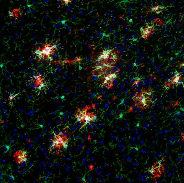
Figure 4. Confocal immunofluorescent analysis of fixed, frozen brain from an amyloid mouse model of Alzheimer's disease using Dectin-1/Clec7a (E3P5W) Horse Chimeric mAb #43191 (gray), Iba1/AIF-1 (E4O4W) Mouse Chimeric mAb (green), β-Amyloid (D54D2) Rabbit mAb #8243 (red), and ProLong Gold Antifade Reagent with DAPI #8961 (blue). The rabbit primary antibody was detected using an anti-rabbit Fc-specific secondary antibody.
#13. GFAP
GFAP is the primary marker of reactive astrocytes and astrogliosis. Increased GFAP expression is a consistent feature in neurodegenerative diseases. It has been observed in HD brain tissue, and its detectability in plasma and CSF makes it a practical complement to examining NfL in biofluid studies.5
- GFAP (D1F4Q) Rabbit mAb #12389 — WB, IHC, IF; H, M, R
- GFAP Matched Antibody Pair #44997 — ELISA+; H, M, R
- PathScan® RP GFAP Sandwich ELISA Kit #56955 — ELISA+; H, M, R
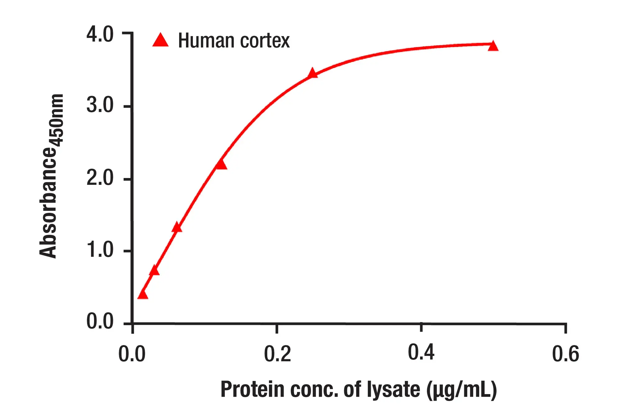 Figure 5. Sandwich ELISA data using GFAP Matched Antibody Pair #44997 are shown. The two antibodies, GFAP (D1F4Q) Rabbit Monoclonal Antibody (BSA and Azide Free) #37608 and GFAP (E4L7M) Rabbit Monoclonal Antibody (BSA and Azide Free) #45946, function as a capture-target-detection sandwich to detect endogenous protein expression in human cortex tissue.
#14. IL-6
IL-6 is a pro-inflammatory cytokine associated with central and peripheral immune activation in HD. Elevated IL-6 has been detected in plasma before motor onset and in CSF from HD gene expansion carriers, supporting its use as a complementary inflammatory readout.7,8
- IL-6 (D5W4V) Rabbit mAb (BSA and Azide Free) #65145 — WB, IF, F; M
#15. YKL-40 (CHI3L1)
YKL-40, also known as CHI3L1, is an inflammation-associated glycoprotein linked to activated microglia and astrocytes. Elevated CSF YKL-40 has been associated with HD stage, clinical severity, and markers of neuronal injury, making it a promising biomarker for connecting neuroinflammation with disease progression8,9
- YKL-40 (E2L1M) Rabbit mAb #47066 — WB, IHC, IF; H
- YKL-40 (E2L1M) Rabbit mAb (BSA and Azide Free) #44073 — WB, IHC, IF; H
Additional Neuroinflammation Targets Worth Exploring
Additional neuroinflammation targets represent a promising avenue for evaluating therapeutic impacts in HD research, and the following biomarkers can help assay developers build broader immune profiling panels:
- CD206/MRC1: A phagocytic microglial phenotype marker useful for distinguishing anti-inflammatory from activated microglial states: CD206/MRC1 (E6T5J) Rabbit mAb #24595 (WB, IP, IHC, IF; H, M, R, Mk) and CD206/MRC1 (E2L9N) Rabbit mAb #91992 (WB, IHC, IF; H)
- CD3: A pan-T cell marker for studying adaptive immune infiltration in HD tissue: CD3 epsilon (D7A6E) Rabbit mAb #85061 (WB, IHC, IF; H, Mk) and CD3 epsilon (D7A6E) Rabbit mAb (BSA and Azide Free) #24581 (WB, IHC, IF; H, Mk)
- Galectin-9: An emerging neuroinflammation target that signals through TIM-3 and has been linked to pro-inflammatory microglial states in neurodegenerative disease models.¹⁰, ¹¹ Ongoing research is investigating this pathway in HD-specific striatal neuroinflammation, making Galectin-9 an emerging target: Galectin-9 (D9R4A) Rabbit mAb #54330 (WB, IHC, F; H) and Galectin-9 (D9R4A) Rabbit mAb (BSA and Azide Free) #71325 (WB, IHC, F; H)
- IL-8: A pro-inflammatory chemokine elevated in plasma and striatal tissue in earlier HD studies.7 Recent work has also found increased IL-8 in serum and HD-affected brain regions, with prominent expression in striatal microglia.12Further research on CSF findings is needed for this exploratory inflammatory target.5,8 IL-8 (E5F5Q) Rabbit mAb #94407 (WB, IHC, IF, F; H) and IL-8 (E5F5Q) Rabbit mAb (BSA and Azide Free) #10681 (WB, IHC, F; H)
- TSPO: a mitochondrial marker used in HD mouse and human PET neuroinflammation imaging; its relationship to microglial activation state is more complex in human tissue than in rodent models: TSPO (D1N7Z) Rabbit Monoclonal Antibody #70358 (WB, IP, IHC, IF; H) and TSPO (F5B8F) Rabbit Monoclonal Antibody #99128 (WB, IP, IHC, IF; H)
Building HD Assays for Biofluid Studies
Biofluid-based measurement is increasingly central to HD research, particularly as clinical trials require sensitive, reproducible detection of mHTT, NfL, and inflammatory markers in CSF and plasma.5, 3, 9 CST antibodies can be formulated without carrier proteins or sodium azide. These BSA- and azide-free formats are ideal for immunoassay development using high-sensitivity platforms such as the Quanterix Simoa and MSD, which enable ultrasensitive detection of low-abundance targets like mHTT in CSF.
To explore Matched Antibody Pairs and ELISA+ assay development resources for neurodegenerative disease research, visit cellsignal.com/research/immunoassay-development. For a broader overview of biofluid tools across neurodegeneration, read our blog, Antibody Toolbox: Neurodegeneration Biomarkers with Biofluid Samples.
CST Antibody Toolkit for Huntington's Disease Research
|
Target |
Product |
Applications |
Reactivity |
|
CD206/MRC1 |
WB IHC IF |
H |
|
|
CD206/MRC1 |
WB IP IHC IF |
H M R Mk |
|
|
CD3 |
WB IHC IF |
H Mk |
|
|
CD3 |
WB IHC IF |
H Mk |
|
|
DARPP-32 |
WB IHC IF |
M R |
|
|
DARPP-32 |
WB |
H M R |
|
|
DARPP-32 |
WB |
H |
|
|
DARPP-32 |
WB |
H M R |
|
|
DARPP-32 |
WB |
H M R |
|
|
Galectin-3 |
WB IHC IF |
H M R |
|
|
Galectin-9 |
WB IHC F |
H |
|
|
Galectin-9 |
WB IHC F |
H |
|
|
GFAP |
WB IHC IF |
H M R |
|
|
GFAP |
ELISA |
H M R |
|
|
GFAP |
ELISA |
H M R |
|
|
GPR52 |
WB IF |
H |
|
|
GPR52 |
WB IF |
H |
|
|
HAP40 |
WB |
H M |
|
|
HIP1 |
WB |
H M R |
|
|
Huntingtin |
WB IHC IF |
H M R |
|
|
Huntingtin |
WB IF |
H M R |
|
|
Huntingtin |
WB IF |
H M R |
|
|
Huntingtin |
ELISA |
H M R |
|
|
Iba1/AIF-1 |
IF |
H M R |
|
|
Iba1/AIF-1 |
WB IHC IF F |
H M R |
|
|
Iba1/AIF-1 |
IF |
H M R |
|
|
Iba1/AIF-1 |
IF |
H M R |
|
|
Iba1/AIF-1 |
IF |
H M R |
|
|
IL-6 |
WB IF F |
M |
|
|
IL-8 |
WB IHC IF F |
H |
|
|
IL-8 |
WB IHC F |
H |
|
|
NeuN |
IF |
H M R |
|
|
NeuN |
WB IHC IF |
H M R Mk |
|
|
NeuN |
IF |
H M R |
|
|
NeuN |
IF |
H M R |
|
|
NeuN |
IF |
H M R |
|
|
Neurofilament-L |
Neurofilament-L (E3V7Y) Rabbit mAb (BSA and Azide Free) 86100 |
WB ELISA |
H M R |
|
Neurofilament-L |
Neurofilament-L (C28E10) Rabbit mAb (BSA and Azide Free) 10768 |
WB IHC IF ELISA |
H M R |
|
Neurofilament-L |
ELISA |
H M R |
|
|
Neurofilament-L |
ELISA |
H M R |
|
|
PGC-1ɑ |
WB |
H M R |
|
|
PSD-95 |
WB IF |
H M R |
|
|
PSD-95 |
IF |
H M R |
|
|
PSD-95 |
IF |
H M R |
|
|
PSD-95 |
IF |
H M R |
|
|
PSD-95 |
IF |
H M R |
|
|
PSD-95 |
WB |
H M R |
|
|
SP1 |
WB IHC IF F ChIP |
H Mk |
|
|
TSPO |
WB IP IHC IF |
H |
|
|
TSPO |
WB IP IHC IF |
M |
|
|
YKL-40 (CHI3L1) |
WB IHC IF |
H |
|
|
YKL-40 (CHI3L1) |
WB IHC IF |
H |
Select References
- MacDonald ME, Ambrose CM, Duyao MP, et al. A novel gene containing a trinucleotide repeat that is expanded and unstable on Huntington's disease chromosomes. Cell. 1993;72(6):971-983.
- Tabrizi SJ, Flower MD, Ross CA, Wild EJ. Huntington disease: New insights into molecular pathogenesis and therapeutic opportunities. Nat Rev Neurol. 2020;16(10):529-546.
- Fanti R, Ayoubi R, Alende C, et al. A guide to selecting high-performing antibodies for Huntingtin (UniProt ID: P42858) for use in western blot, immunoprecipitation, and immunofluorescence. F1000Res. 2025;13:922.
- Parsons MP, Kang R, Buren C, et al. Bidirectional control of postsynaptic density-95 (PSD-95) clustering by Huntingtin. J Biol Chem. 2014;289(6):3518-3528.
- Paulsen JS, Bovin NA, Clemsen JD, et al. Systematic review with meta-analysis of biofluid markers for Huntington's disease. Mov Disord. 2025;40(12):2578-2595.
- Siew JJ, Chen HM, Chen HY, et al. Galectin-3 is required for the microglia-mediated brain inflammation in a model of Huntington's disease. Nat Commun. 2019;10(1):3473.
- Björkqvist M, Wild EJ, Thiele J, et al. A novel pathogenic pathway of immune activation detectable before clinical onset in Huntington's disease. J Exp Med. 2008;205(8):1869-1877.
- Rodrigues FB, Byrne LM, McColgan P, et al. Cerebrospinal fluid inflammatory biomarkers reflect clinical severity in Huntington's disease. PLoS One. 2016;11(9):e0163479.
- Bondulich MK, Phillips J, Cañibano-Pico M, et al. Translatable plasma and CSF biomarkers for use in mouse models of Huntington's disease. Brain Commun. 2024;6(1):fcae030.
- Peng Q, Zhang G, Guo X, et al. Galectin-9/Tim-3 pathway mediates dopaminergic neurodegeneration in MPTP-induced mouse model of Parkinson's disease. Front Mol Neurosci. 2022;15:1046992.
- Guo X, Zhang G, Peng Q, et al. Galectin-9 activates the TLR4-NLRP3 inflammasome pathway and promotes tau pathology in Alzheimer's disease. Brain Behav Immun. Published online October 26, 2025.
- Fung JN, Lee JD, Adam R, O'Sullivan JD, Woodruff TM. Peripheral and central elevation of IL-8 in patients with Huntington's disease. Mol Immunol. 2025;179:84-93.
- Rocha NP, Ribeiro FM, Furr-Stimming E, Teixeira AL. Microglia activation in basal ganglia is a late event in Huntington disease pathophysiology. Neurol Neuroimmunol Neuroinflamm. 2021;8(3):e984.
NDG-93768