True innovation comes from collaboration–from bringing experts together to create techniques and tools that solve challenges in new ways. The spatial biology revolution is one example of how combining tools and talent brings about forward progress. Named Method of the Year by Nature in 2020, spatial biology melds a combination of technologies including “concepts and technology from microarrays, imaging, sequencing, and bioinformatics analysis” to provide data and positional context of cells in tissue. A rapidly evolving field, new tools, techniques, and methods continue to be developed to speed spatial proteomics research.
That’s why we’re so excited about our partnership with Leica Microsystems (LMS) to validate antibodies for the Cell DIVE Multiplexed Imaging Solution. Through the collaboration, we’re bringing together antibody experts from CST and imaging experts from LMS to help scientists accelerate spatial biology research by removing one of the most time-consuming and cost- and labor-intensive parts of the research process: Antibody panel validation.
As part of this project, Reggie Prioli, PhD, formerly the head of our antibody conjugation group, along with CST scientist Emily Alonzo, PhD, and others, worked with LMS scientist Melinda Hill, PhD to identify and validate 30 antibodies essential for oncology research, which will join the 75 CST antibodies that have already been validated by LMS for use on Cell DIVE.
This post provides an inside look at the partnership, the team’s antibody selection and validation process, and what’s to come next.
Spatial Biology Research on Cell DIVE: Multiplex 60+ Biomarkers
Powerful new imaging and computational tools like Cell DIVE enable the interrogation of normal and diseased cells within their spatial contexts. This is helping to further elucidate the biological basis of disease and answer important questions such as why one patient may respond positively to a therapy while another does not.
 “[However], the traditional approach for tissue imaging is an iterative, serial process,” explains Rick Heil-Chapdelaine, Cell DIVE Sales Specialist at LMS. “To get more markers, you have to use multiple tissue sections from a sample. What you end up with is an understanding of the cellular neighborhoods, but not the individual cells within that neighborhood, since you get different cells in each section.”
Cell DIVE, on the other hand, can multiplex 60+ biomarkers within a single tissue sample using an iterative technique that includes biomarker staining, sample imaging, and a gentle chemical dye inactivation process. Essentially, if you think of antibodies and associated fluorophores as lightbulbs, the dye inactivation process allows researchers to turn the lights off after imaging–but leave the lightbulbs in place. Additional lightbulbs can be added, imaged, and switched off, without negatively affecting the tissue sample (Figure 1).
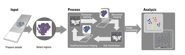
Figure 1: The Cell DIVE iterative multiplexing process. Image courtesy of Leica Microsystems.
“What Cell DIVE allows us to do is take a single sample and go through an iterative process of staining then dye inactivation, staining then dye inactivation, staining then dye inactivation, to produce crystal-clear images that are stitched together for downstream statistical analysis,” Rick explains. This process is illustrated in Figure 2.
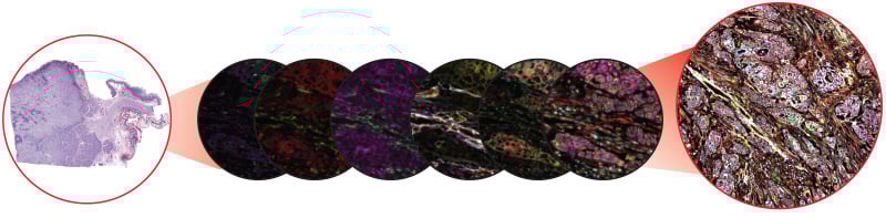 Figure 2: The Cell DIVE Multiplexed Imaging Solution can produce crystal-clear images of whole tissue using 60+ biomarkers, with automatic calibration and correction to enable robust downstream analysis. The figure above shows six imaging rounds with four biomarkers per round (center), as well as the final stitched image showing 24 biomarkers (right). Image courtesy of Leica Microsystems.
One of the most unique aspects of Cell DIVE is that slides can be stored away after imaging and then re-stained for additional biomarkers months or even years later.
“You can store your slides and return to them when your hypothesis evolves or new biomarker targets are released to further study the same tissue sample,” explains Melinda. “Instead of re-staining for all the biomarkers you’ve already tested for, you can overlay the original slide with additional biomarkers to probe for more data in the original context. This can help speed discoveries and is a time- and money-saver.”
However, while the Cell DIVE offers flexibility in antibody selection and custom antibody panel design, antibodies used on the device need to be validated and verified for instrument compatibility–in addition to specificity and sensitivity.
“The antibodies are really at the heart of the whole Cell DIVE process,” says Rick. “And having quality antibodies that are both specific and sensitive enough to work on the Cell DIVE system is key for having a downstream analysis that is trustworthy.”
Multiplexing You Can Count On: Antibody Selection and Validation for Cell DIVE
The ongoing reproducibility crisis in the scientific community means that researchers must be extra careful when selecting and validating antibodies for use in their experiments. LMS has tested around 2,000 commercially-available antibodies, but only about 350 are verified for use on the Cell DIVE system. This means that about two in three antibodies failed either due to sensitivity, specificity, or both. Additionally, only about 10% of the validated antibodies are available from manufacturers as commercial conjugates, meaning that researchers have to perform conjugation themselves in order to use the antibodies on the Cell DIVE.
“The reason there are such low validation numbers is because the majority of antibodies from vendors can be highly variable in quality,” explains Melinda. “That's why high-quality antibodies are essential and why we’re so excited about this ongoing collaboration. CST is the leader for high-quality, validated antibodies, and we’ve found that as long as the CST antibody is validated to work in IHC and FFPE, we can almost always trust that they will work with Cell DIVE.”
The only variable that remains is conjugation. For an antibody to work on the Cell DIVE, it must be able to be conjugated to a fluorophore to enable the instrument’s iterative process. The degree of labeling (DOL) is also important, as well as the antibody concentration, both of which must be optimized in order to achieve success on the Cell DIVE imager. Therefore, having commercially available conjugates created by conjugation experts can be critical for achieving successful imaging.
So, how were CST antibodies selected and verified for use on the Cell DIVE Multiplexed Imaging Solution?
Antibody Selection
At the outset of the LMS and CST partnership, the team prioritized antibodies based on their usefulness for spatial biology research, their applicability to specific disease states, and the likelihood that the antibody would work on the system.
“When dealing with spatial biology within tissues, there are three types of antibody markers you want to look at: ones that tell us where the cells are, one that tells us what they are, and ones that tell us what they’re doing,” explains Rick. “After that, you can start looking at the spatial information, such as ‘What is it about our neighbors that is influencing a particular response?’”
The team’s initial list of antibodies followed this general rule and included antibodies used for segmentation and phenotyping, especially oncology markers used to help understand how the tumor microenvironment (TME) evolves during tumorigenesis and treatment response. Immune cell markers for interrogating myeloid and lymphoid cell lineages and structures were prioritized, as well as those used to detect key proteins in the TME, such as Vimentin, Ki-67, TIM-3, and CD45. Antibodies that are available as CST® conjugates or as custom conjugations were also prioritized.
The final list included about 30 CST antibodies.
Antibody Validation
To validate the antibodies, the team examined the expression of the biomarkers across 12 different human cancer tissues, including both whole tissue and tissue microarray (TMA) slides imaged on Cell DIVE. The validation study looked at the expression of tumor and immune cell lineages in the TME, and captured their spatial relationships.
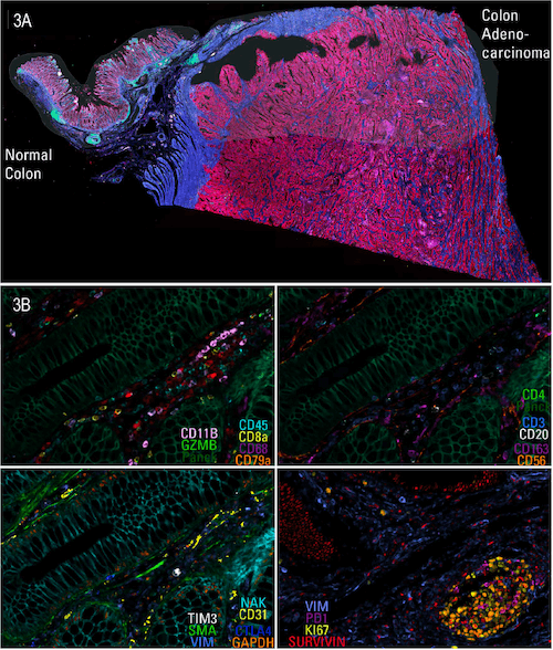 Figure 3: Multiplexed imaging of the CST antibody panels enables an examination of immune cell components in colon adenocarcinoma (CAC) tissue on a single slide.
Ten different testing rounds were conducted to ensure continued affinity after dye inactivation. The biomarker antibodies were conjugated and randomly assigned to a staining round without optimization. After each step, antibodies were compared to the literature and each other and evaluated for specificity and sensitivity.
The resulting data panel enables the identification of clusters containing cells of different immune classes, cell types, and subtypes and provides unprecedented and novel insights into immune cell populations, among other cell populations, and spatial cell interactions in the TME.
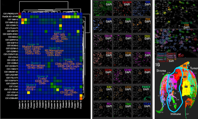 Figure 4: Cluster analysis following segmentation using 30 biomarkers reveals the immune cells within the tissue and their relationship to other immune cells and cell clusters (UMAP).
“Based on the fail rate of our prior validations, you would have expected about one in three antibodies to fail—so about ten to twelve on our initial list of 35,” explains Melinda. “Instead, we found that 33 out of the 35 worked on the Cell DIVE, which speaks to the high-quality nature of the CST antibodies and conjugations.”
The final project was summarized in the poster presentation, Capturing the Spatial Landscape of Tumor and Immune Cell Lineages in the Microenvironment of Human Cancer Tissues, which was presented at the Society for Immunotherapy of Cancer (SITC) annual meeting in 2022.

Validated by LMS and CST—So You Don’t Have to
With this partnership, the CST and LMS teams hope to take some of the guesswork out of antibody selection for multiplex imaging and save researchers precious time and frustration.
“We asked our customers what words came to mind when they thought about the antibody conjugation process and we got some pretty interesting responses,” concludes Rick. “Some of the most common answers were things like ‘time-consuming,’ ‘complicated’—so we know many scientists aren’t excited about it.”
By bringing together microscope and imaging experts from LMS and antibody experts from CST, this collaboration helps provide researchers not only with the right tools for conducting spatial biology research, but also the ability to trust that the tools will work. This will allow researchers to spend more time doing what they do best—discovering novel ways to explore the mechanisms of disease.
“What we’re excited about is being able to do some of this work for researchers so that we can reduce these hurdles for them,” says CST scientist Emily Alonzo. “This will let them get to the real experiment faster, so they can get at the data they’re truly interested in.”
In the year ahead and as part of this ongoing partnership, the team will continue to validate more antibodies for use on the Cell DIVE imager, so be sure to check back for updates!
Additional Resources:
- Learn more about how CST validates antibodies for IHC.
- Read the partnership press release.
22-BPA-51810