


Host-based multiplex immunofluorescence (IF) of fixed frozen brain tissue is a powerful approach for visualizing multiple cellular targets within the same tissue section. However, expanding the number of targets has long depended on finding primary antibodies from diverse host species—a strategy limited by the availability, specificity, and performance of non-rabbit reagents.
Chimeric antibodies are powerful reagents that unlock flexible multiplex IF panel design while preserving data quality. Manufactured in vitro using entirely animal-free gene cloning, CST® chimerics are recombinant monoclonals in which the binding domain of a highly specific rabbit antibody has been engineered with the backbone of a different host species—either horse, mouse, or feline.
 The result is a trusted portfolio of flexible reagents that work within your existing IF protocols. You can use CST chimerics to build neuro-focused panels targeting key neuronal, microglial, astrocyte, and oligodendrocyte markers—expanding your multiplex capabilities while preserving data quality.
Ready to design your own experiment? Browse CST Chimeric antibodies.
How to Design a Multiplex IF Experiment Using Chimeric Antibodies
CST chimeric antibodies contain the same binding domain as their parental source. This means they have the same binding profile as their parent antibody. They are ideal for low-plex (three to four target) immunofluorescence with fixed frozen brain tissue using the standard fixation, permeabilization, and incubation workflows you're familiar with.
Neuronal & Glial Cell Characterization
Chimeric antibodies can be used to easily profile neurons, astrocytes, and microglia on the same section. For example, you can use a mouse chimeric Beta3-tubulin antibody to label neurons, a rabbit anti-GFAP antibody to visualize astrocytes, and a horse chimeric Iba1 antibody for microglia in the same slide during the primary incubation step./70668_B3-Tubulin%20Mouse%20Chimeric%20Antibody.png?width=520&height=350&name=70668_B3-Tubulin%20Mouse%20Chimeric%20Antibody.png)
Detection of neuronal and glial cells in mouse brain tissue. IF analysis of fixed frozen mouse cerebellum using beta3-Tubulin (D71G9) Mouse Chimeric Monoclonal Antibody #70668 (green), Iba1/AIF-1 (E4O4W) Horse Chimeric Monoclonal Antibody #23757 (gray), GFAP (E4L7M) Rabbit Monoclonal Antibody #80788 (red), and ProLong Gold Antifade Reagent with DAPI #8961 (blue). Rabbit primary antibody is detected using an anti-rabbit Fc-specific secondary antibody.
For clear, specific detection, use species-specific secondary antibodies conjugated to distinct fluorophores to enable flexible signal separation and minimize cross-reactivity. You can combine these secondaries during a single incubation step to streamline your workflow. Because CST chimeric antibodies preserve their rabbit-derived binding domains, they remain fully compatible with any unconjugated rabbit antibodies or existing reagents you already rely on in your lab, with minimal concern for cross-reactivity. When your chimeric antibody shares a host species with another primary, be sure to use an Fc-directed secondary antibody to ensure accurate detection.
By selecting chimeric antibodies for your key cell-type markers, you can streamline panel creation and ensure species compatibility in your experimental design.

Detecting Amyloid Plaques & Classifying Surrounding Cell State Pathology
Chimeric antibodies can also be used to uncover cell-type specificity by multiplexing markers around amyloid plaques to identify pathological cellular subtypes.
For example, in an amyloid mouse model of Alzheimer’s disease, you can combine a Beta-amyloid rabbit monoclonal to identify amyloid plaques, a chimeric mouse Iba1 antibody to detect all microglia, and a horse chimeric Dectin-1/Clec7A antibody for disease-associated microglia.
/2025_SfN_Chimeras-1.webp?width=350&height=350&name=2025_SfN_Chimeras-1.webp)
Detection of amyloid plaques and surrounding cells. IF analysis of fixed frozen brain from an amyloid mouse model of Alzheimer's disease using Iba1 (E4O4W) Mouse Chimeric Monoclonal Antibody #58410 (green), beta-Amyloid (D54D2) Rabbit Monoclonal Antibody #8243 (red), Dectin-1/Clec7a (E3P5W) Horse Chimeric Monoclonal Antibody #43191 (gray), and ProLong Gold Antifade Reagent with DAPI #8961 (blue).
Mapping Functional Neuronal Activity
For functional neuron studies, chimeric antibodies enable high-fidelity detection of active populations.
For example, combining a chimeric mouse Tyrosine Hydroxylase antibody with a rabbit NeuN marker and a chimeric feline c-Fos antibody provides a neuronal activity readout mapping dopaminergic neuron activation in adult wild-type mice. The result is a multi-target view of neuron identity, morphology, and functional state within a single imaging workflow.
/42157_neuronal%20activity%20chimerics.png?width=350&height=350&name=42157_neuronal%20activity%20chimerics.png)
Detection of neuronal activity in dopaminergic neurons. IF analysis of fixed frozen mouse olfactory bulb using NeuN (D4G4O) Rabbit Monoclonal Antibody #24307 (green), Tyrosine Hydroxylase (E2L6M) Mouse Chimeric Monoclonal Antibody #29085 (red), c-Fos (E2I7R) Feline Chimeric Monoclonal Antibody #42157 (gray), and ProLong Gold Antifade Reagent with DAPI #8961 (blue).
Chimeric antibody technology simplifies multiplex immunofluorescence by converting rigorously characterized rabbit monoclonal antibodies into a versatile collection of off-species reagents. This strategy removes the challenge of sourcing compatible primary antibodies, enabling a straightforward workflow for clear and reliable multi-target detection in order to answer more complex biological questions.
Experimental Considerations and Optimization Tips
As with any IF experiment, tissue sections and cultured cells demand different processing workflows, and careful sample preparation and antibody validation are crucial. Always confirm that an antibody is validated for your species of interest and review the Sensitivity and Specificity information on the product datasheet.
- Concentration: Primary antibody binding is concentration-, time-, and temperature-dependent, regardless of source (rabbit or chimeric). The CST product datasheet provides a starting dilution range, but optimal conditions often require titration for the specific sample type. Too high a concentration can increase background, while too low a concentration yields weak or no signal.
- Incubation: Incubation time affects signal intensity—longer, colder incubations often improve specificity, which is why we recommend incubating overnight at 4°C.
- Secondary Antibody Selection: Chimeric antibody staining is a type of indirect IF, therefore requiring a secondary antibody. Critical elements to consider here are the correct host species reactivity, compatibility of the fluorophore (dye) with the microscope's filters and lasers (excitation/emission spectra), and incubation conditions. Using the wrong secondary, too high a concentration, or omitting a secondary-only negative control are common pitfalls.
Simplify your experimental design by choosing validated secondaries from CST.
Simplify Your Multiplex IF: More Hosts, Same Reliable Performance
Extensive validation testing shows that CST chimeric antibodies perform identically to their parent rabbit monoclonal antibodies—so you can trust them to deliver the same high specificity and sensitivity in your experiments.
By redesigning these trusted rabbit monoclonals into horse, mouse, or feline isotypes, CST chimeric antibodies make it easy for you to expand your multiplex panels without changing your existing workflow or compromising data quality. This approach simplifies experimental design and execution, enabling clean, crosstalk-free detection of multiple neuronal, glial, and disease markers within a single section.
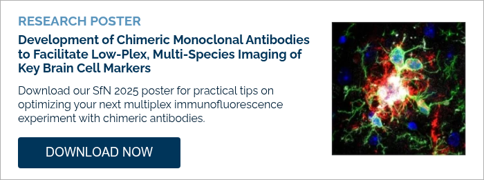
Want to learn more about setting up a multiplex IF experiment for your next neuro project? Download our SfN 2025 poster to learn more.
Additional Resources
- Resource Center: Multiplex Solutions for Spatial Biology
- Blog: Anything but Rabbit: Why Multiplex with Chimeric Antibodies?
- Blog: Strategies for Successful Multiplex IF Experiments Using Conjugated Antibodies


