


You've just finished running your lysate sample by SDS-PAGE, so you're pretty confident your protein should have migrated at the right molecular weight and is ready to transfer to a membrane for detection by western blot (WB). But what if it's not?
If, after going through the full protocol, your band isn't where you expect it, you may need to troubleshoot your antibodies, but you should also troubleshoot the electrophoresis and transfer steps. This can be done by visualizing total protein by staining with Ponceau S or Coomassie.
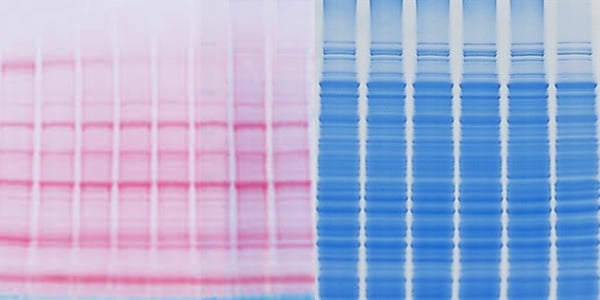
Ponceau S Staining Solution #59803 and Coomassie Brilliant Blue Stain allow for the visualization of protein transfers after electrophoresis. They are important for confirming protein transfer and presence of the target of interest, saving time and valuable resources in your experiments. Additionally, they show any imperfections in the blot such as bubbles and uneven transfer, which are important considerations when aiming for publication-quality images.
Both Ponceau S and Coomassie Brilliant Blue stains are negatively charged solutions that bind to positively charged amino acid groups and non-polar protein regions. Ponceau S detects protein levels at 200 ng and higher, and is compatible with PVDF, nitrocellulose, or nylon membranes. The traditional Coomassie Brilliant Blue is only compatible with PVDF membranes, but can detect protein levels at 50 ng and higher. A detection stain with higher sensitivity can be important, depending on the type of sample and testing being done. However, the use of Coomassie Brilliant Blue solution prevents the possibility of continuing on with WB analysis, as the alcohols and acids present in the solution cause the fixation of the protein samples within the gel after use.
Coomassie Blue G-250 Staining Protocol
There are several types of Coomassie Brilliant Blue staining solutions available, and protocols vary slightly, taking anywhere from two hours to a full day, depending on the vendor and solution type. One of the most commonly used Coomassie Blue G-250 can be used as follows:
- After electrotransfer, wash the polyacrylamide gel three times with ddH2O.
- Add at least 25mL (or until the gel is covered) of the staining solution onto the polyacrylamide gel.
- Place the gel with the staining solution onto a shaker and agitate slowly to prevent gel from adhering to the container.
- The staining time varies depending on gel thickness and the percentage of acrylamide. It can take two hours or more to see a uniform blue color on the gel.
- After the gel has been stained, wash the gel with ddH2O and gently shake for one to two hours, changing the ddH2O several times in between to remove the excess free dye.
Ponceau S Staining Protocol
The Ponceau S staining protocol takes about 20 minutes, is non-toxic, and is a gentler solution than Coomassie Brilliant Blue. Ponceau S staining solution does not fix the protein, allowing for WB analysis after staining, which is another key consideration.
To use Ponceau S (CST #59803):
- Following electrotransfer onto the membrane, briefly rinse the membrane in ddH2O.
- Incubate the membrane in Ponceau S staining solution for five to ten minutes at room temperature.
- Wash the membrane in ddH2O for one to five minutes, until red bands are visible.
- Bands can then be marked or photographed prior to rinsing the membrane with a few washes of 1X TBS-T for five minutes.
The experiment can now continue, and any remnants of Ponceau S will be washed away during the blocking step. Furthermore, Ponceau S does not interact with antibody binding.
When to Use Ponceau S vs Coomassie Brilliant Blue
Overall, both solutions can help to guarantee your results. If you need to detect amounts of protein in the 50-200 ng range, Coomassie Blue is your best option.
However, at Cell Signaling Technology, Ponceau is often our top choice because of its speed, ease of use, and gentle but effective nature.
Select References
- Chevalier F. Standard Dyes for Total Protein Staining in Gel-Based Proteomic Analysis. Materials (Basel). 2010;3(10):4784-4792. Published 2010 Oct 20. doi:10.3390/ma3104784
- Salinovich O, Montelaro RC. Reversible staining and peptide mapping of proteins transferred to nitrocellulose after separation by sodium dodecylsulfate-polyacrylamide gel electrophoresis. Anal Biochem. 1986;156(2):341-347. doi:10.1016/0003-2697(86)90263-0
- Seeing blue and red: Coomassie vs. Ponceau Staining. Advansta Inc. https://advansta.com/seeing-blue-red-coomassie-vs-ponceau-staining/. Published March 22, 2016.
- Goldman A, Harper S, Speicher DW. Detection of Proteins on Blot Membranes. Curr Protoc Protein Sci. 2016;86:10.8.1-10.8.11. Published 2016 Nov 1. doi:10.1002/cpps.15


