Guest blogger Charles Haitjema, PhD, is a Research Area Manager at Bio-Techne. This post details the CeST antibodies that have been validated for use on Simple WesternTM systems from ProteinSimpleTM, a Bio-Techne brand.
—
We are thrilled that Simple Western has been added as a validated application to the catalog of high-quality antibodies from CST. Now, Simple Western users can easily find validated antibodies by filtering for Simple Western as an application on the CST website—with hundreds of antibodies already validated and more being added all the time.
What is Simple Western?
Simple Western is a capillary electrophoresis immunoassay that is automated inside of a benchtop instrument like Jess™, Abby™, Peggy Sue™, Sally Sue™, and NanoPro™ 1000 (Figure 1). As an open immunoassay platform, Simple Western users may choose any commercial or custom antibody for specific detection of target proteins and protein isoforms. Since the first automated western blot instrument was introduced in 2009, Simple Western users have relied heavily on the extensive catalog of high-quality primary antibodies from CST to develop their assays.
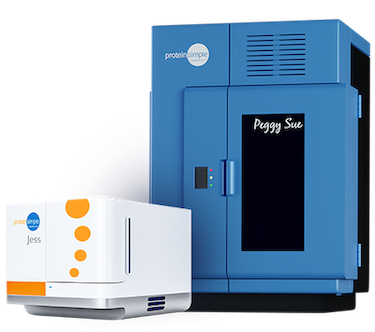
Figure 1. Simple Western instruments Jess (left) and Peggy Sue (right). Jess performs size-based capillary electrophoretic immunoassays with multi-channel multiplexing in chemiluminescence and NIR/IR fluorescence channels. Peggy Sue performs size or charge-based capillary electrophoretic immunoassays with chemiluminescence detection. Jess processes up to 25 samples in three hours, and Peggy Sue processes up to 96 samples overnight. Image courtesy of ProteinSimple.
CST® Antibodies Validated for Simple Western
CST is a company founded by scientists for scientists, with a long history in signaling. Ensuring that antibodies are stringently validated for use in each application type is one of the company’s core tenets, and the reason why CST has been a go-to supplier of WB antibodies for decades—and was named the CiteAb Antibody Supplier of the Decade.
The CST antibodies validated for Simple Western allow you to skip the optimization steps of determining the best assay conditions for your experiment. Product scientists from CST and Simple Western experts have already performed this step for you, so you know the ideal dilution factor to get the best results before starting your experiment.
You can find everything you need to get up and running in the product datasheet and product webpage for each of the Simple Western-validated antibodies:
Simple Western Overcomes Limitations of Immunoassays Like Western Blot and ELISA
As its name suggests, Simple Western performs western blot-like analysis and is the only platform on the market that provides a fully automated western blotting workflow, offering high reproducibility, sensitivity, and quantitation, in addition to time and cost savings.
So, what is the difference between western blot and Simple Western? To begin with, Simple Western is fully automated following sample preparation and plate loading. The manual steps of traditional western blot are precisely controlled inside of an automated capillary benchtop instrument. Instead of SDS-PAGE gels, Simple Western uses capillary electrophoresis (CE) to separate proteins by molecular weight (size) using CE-SDS or by isoelectric point (charge) using cIEF (Figure 2).
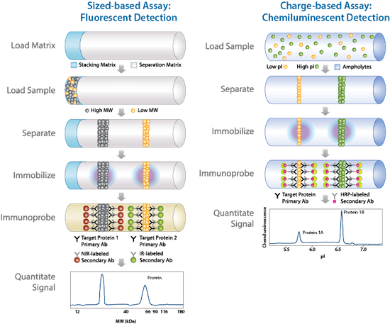 Figure 2. Simple Western performs immunoassay analysis inside of capillary. Samples are separated by size using CE-SDS (left panel) or by charge using cIEF (right panel). All steps of matrix and sample load, separation, immunoprobe, and signal quantitation are automated in a precisely controlled capillary environment. Image courtesy of ProteinSimple.
Fully Quantitative Data with Reproducibility You Won’t Get with Traditional Western Blot
Once protein separation is complete, proteins are covalently immobilized to the capillary wall, and the steps of traditional western blot like washing, antibody incubation, and immunodetection are seamlessly performed back-to-back in the capillary without user intervention (Figure 2). Therefore, Simple Western eliminates the gel-to-membrane transfer of traditional western blots. Once the run is complete, data automatically appears in the Compass for Simple Western software. By automating the steps of traditional western blots inside of a precisely controlled capillary environment, Simple Western produces fully quantitative, highly reproducible data with up to 6-log dynamic range and CVs consistently below 15%.
The advantages do not stop there. Compared to western blot, Simple Western requires only a fraction of sample volume, as little as 3 µL, and the Jess and Abby instruments process 25 samples in as little as three hours. Peggy Sue, Sally Sue, and NanoPro 1000 process 96 samples overnight and can generate up to eight data points per 5 µL of sample. Jess users can choose from flexible multiplex detection strategies in chemiluminescence and NIR/IR fluorescence channels (Figure 3). Stellar™ NIR/IR Modules for Jess provide industry-leading detection sensitivity that is 100X more sensitive than the closest competing fluorescence western blot imager. Looking for accurate protein expression measurements? Simple Western provides simultaneous total protein detection (Figure 3), eliminating the use of unreliable housekeeping proteins for data normalization and allowing for more accurate protein expression measurements.
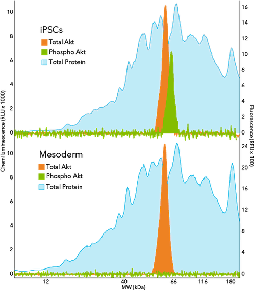 Figure 3. Simple Western analysis of total Akt, phospho Akt, and total protein across undifferentiated iPSCs and iPSC-derived mesodermal cells using the Stellar assay on Jess. CST antibodies targeting total AKT (#58295) and phosphorylated Akt (#9271) isoforms were detected using the Stellar NIR/IR Modules, and total protein was simultaneously detected using the Stellar Total Protein Module in the chemiluminescence channel. Image courtesy of ProteinSimple.
Simple Western even eliminates the strip and re-probe method of traditional western blot with the RePlex™ feature on Jess and Abby. RePlex performs sequential immunoassays back-to-back in the same capillary so that more targets can be probed in the same sample. Because samples are covalently immobilized to the capillary wall, RePlex completely and reproducibly removes antibodies between probing cycles. So, unlike the strip and re-probe method of western blot, RePlex assays are not impacted by signal interference or sample loss between probing cycles. The second cycle may also be used for total protein detection to normalize protein expression data (Figure 4).
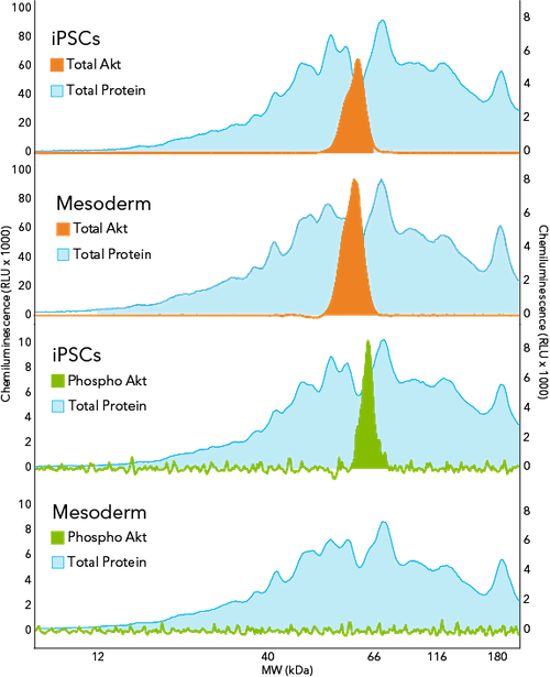 Figure 4. Simple Western analysis of total Akt, phospho Akt, and total protein across undifferentiated iPSCs and iPSC-derived mesodermal cells using the RePlex assay on Abby. CST antibodies targeting total AKT (#58295) and phosphorylated Akt (#9271) isoforms were used in Probe 1 of RePlex and total protein was detected in Probe 2 of RePlex. Image courtesy of ProteinSimple.
Simple Western Versus ELISA: Less Matrix Effect, Simpler Assay Development, Plus Protein Separation
When it comes to analyzing complex sample types like tissue homogenates, ELISA users often encounter interference from the sample matrix, called matrix effect, which skew results. By contrast, Simple Western uses proprietary chemistry to bind sample analytes to the capillary wall after separation. This step covalently binds even within a complex matrix, reducing the matrix effect that can occur when sample analytes are captured using a specific capture antibody in an ELISA. Following covalent sample immobilization, Simple Western flushes the sample matrix from the capillary before immunodetection in an optimized buffer environment, further reducing matrix effect.
In addition, custom development of sandwich ELISAs can be challenging, owing to the need to identify and validate a pair of two antibodies rather than just a single antibody reactive against the target of interest. Simple Western only needs one target-specific antibody to give specific detection, which simplifies assay development and accelerates development time compared to custom ELISA development. Furthermore, size or charge separation on Simple Western, combined with the specificity of immunodetection, allow you to discriminate between different isoforms of target proteins with high resolution (Figure 5). In a direct comparison to a leading commercial ELISA kit for human eNOS detection in brain tissue homogenate samples, Simple Western outperformed in assay range and sensitivity while ELISA seemed to underestimate eNOS concentration compared to Simple Western (Table 1).
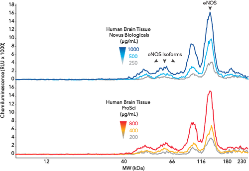
Figure 5. Simple Western analysis of human brain tissue homogenates using a CST antibody targeting Human eNOS (#5880). Image courtesy of ProteinSimple.

Table 1. Comparison of ELISA and Simple Western assays for eNOS quantification in brain tissue.
Expanding Simple Western’s Potential with Cell Signaling Technology
Simple Western is a proven technology that has advanced research and development workflows in a wide variety of applications including cell and gene therapy, cancer and immuno-oncology, protein bioprocessing, cell signaling and signal transduction analysis, targeted protein degradation, vaccine development, neuroscience and neurodegenerative disease, infectious disease, and more.
The large catalog of high-quality CST antibodies has been instrumental in the success of Simple Western—close to 500 Simple Western publications feature antibodies from CST, which represents approximately 25% of all Simple Western publications. With Simple Western added as an application to the CST catalog, Simple Western users can optimize their assay development faster.
As part of this partnership, more modification-state-specific antibodies for studying important molecular signaling pathways will continue to be validated for Simple Western, so be sure to check back for additional information using the link below:
Explore the Simple Western-validated antibodies.
Additional Resources:
- Read the press release to learn more about the partnership between CST and Bio-Techne.
- Learn more about the CST Antibody Validation Principles.
- Learn more about Simple Western.
Cell Signaling Technology and CST are registered trademarks of Cell Signaling Technology, Inc. ProteinSimple, Simple Western, Jess, Abby, Peggy Sue, Sally Sue, NanoPro, Stellar, and RePlex are trademarks of ProteinSimple Corporation. All other trademarks are the property of their respective owners. Visit cellsignal.com/trademarks for more information.
23-BPA-71850


