The death of a cell may be a more complex process than one might initially imagine. Cells expire in many different ways for different reasons. They can die either during development, or as a result of stressful events, such as metabolic disruption, pathogenic invasion, or physiological tissue damage.
Types Cell Death: Type I, II & III
Decades of research have allowed for the detailed characterization and classification of the myriad ways that cells can self-destruct. The demise of a cell can sometimes be caused by rapid overwhelming external influences, as would be the case with sudden physiological injury or accidental cell death (ACD). Alternatively, cell death can occur via dedicated molecular pathways that may be controlled either through pharmacologic or genetic means, and this is referred to as regulated cell death (RCD). Classically, cell death is categorized into three different types, based on morphological changes, triggers, and the biochemical pathways involved.
Apoptosis, or Type I cell death, is a tightly regulated form of programmed cell death (PCD), that triggers cells to self-destruct without any external influence. It is an essential part of life, particularly for multicellular organisms that must control the growth, development, and turnover of cells in order to maintain homeostasis. Apoptosis is critical for proper embryonic development, and a classic example of this process can be observed when cells between the digits on a hand apoptose in order to separate the fingers.

Autophagy, or Type II cell death, is also often categorized as a type of programmed cell death.
However, over time it has become clear that it can promote cell survival as well as cell death, depending upon the circumstance. Autophagy is a process by which cellular organelles and other contents are devoured by lysosomes to clear away unnecessary or dysfunctional components. This critical mechanism allows for the systematic degradation and recycling of cellular materials.
Necrosis, or Type III cell death, has historically been thought of as a process that occurs accidentally due to extreme external physiological stress or ACD. However, in recent years, researchers have shed light on specific regulated molecular mechanisms that can be triggered when a cell is under stress and alternative methods of cell death are not available. Some of these regulated necrotic cell death pathways include necroptosis, pyroptosis, ferroptosis, and NETosis.

Apoptosis
Apoptosis is a tightly controlled pattern of programmed cell death characterized by distinct morphological changes along with the activation of specific caspases and mitochondrial control pathways. It can be triggered via intrinsic or extrinsic pathways. The intrinsic pathway can be activated by cell stress, DNA damage, developmental cues, or withdrawal of survival factors. The extrinsic pathway is triggered via the detection of extracellular death signals from other cells.
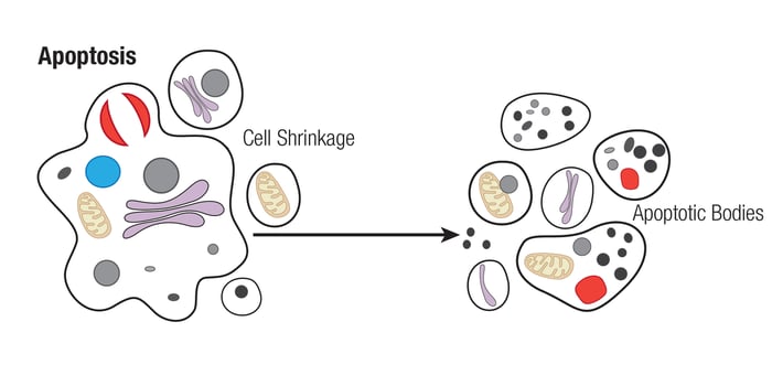
An apoptotic cell can be identified by distinguishing morphological characteristics. It appears shrunken and displays a characteristically deformed membrane referred to as membrane blebbing. It is also characterized by chromatin condensation and nuclear fragmentation. The TUNEL assay uses this DNA fragmentation to identify apoptotic cells and tissue by labeling the 3’OH end of the fragmented DNA with a modified dUTP conjugated to a fluorophore. TUNEL assays are a robust, widely used method to detect apoptotic cells since they do not depend on transient markers. They are also compatible with antibody co-labeling protocols to allow for easy multiplexing with immunohistochemistry, flow cytometry, and immunofluorescence applications. CST offers three TUNEL kits that are simple and easy to use, and deliver same-day results in cultured cells, tissue, and paraffin-embedded samples.
Membrane blebbing can lead to the formation of small vesicles, termed apoptotic bodies, around the edge of the dying cell. It is important to note that the cell membrane remains intact during apoptosis. Since the cell membrane retains integrity during apoptosis, intracellular cytokines and digestive enzymes are prevented from being released into the extracellular space, which limits neighboring tissue damage and inflammation. However, phosphatidylserine lipids within the cell membrane, which normally face the cytosol, flip to become exposed to the extracellular space. Their presence acts as a signal for phagocytes to remove the dead cell. Due to its ability to bind to phosphatidylserine lipids on the cell membrane, Annexin V is often used as a marker of early apoptosis in combination with reagents that confirm the integrity of the cell membrane, such as propidium iodide.
The intrinsic apoptotic pathway involves many conserved signaling proteins and depends upon the integrity of the mitochondria. This process is tightly regulated by a balance between the activity of proteins in the Bcl-2 family, which consists of pro-apoptotic (Bax, Bak, Bad, Bid, Puma, Bim, and Noxa) and anti-apoptotic (Bcl-2, Bcl-xL, Bcl-w, Mcl-1) factors. Apoptosis is triggered when pro-apoptotic family members with a BH3 domain, such as BAD, BID, and BIM, are activated during cellular stress via changes in expression or through post-translational modifications. Then pro-apoptotic proteins, BAX and BAK, induce changes in the mitochondrial outer membrane permeability (MOMP) leading to the release of cytochrome c from the organelle’s intermembrane space. Free cytochrome c then forms a complex with Apaf-1 called the apoptosome, which activates caspase-9 and triggers a cascade of apoptotic events.
Anti-apoptotic family members, such as BCL-2, BCL-xL, and MCL-1, can bind to and inhibit pro-apoptotic proteins to prevent apoptosis. These proteins are often active in cancer cells and are therefore attractive targets for the development of cancer therapeutics. Translational research has focused on targeting these proto-oncogenic proteins in different ways; one strategy has been to design compounds to mimic BH3-containing pro-apoptotic proteins that induce apoptosis by competing with the interaction between pro- and anti-apoptotic family members.
Both the intrinsic and extrinsic pathways are ultimately dependent upon the protease activity of specific members of the caspase family. These caspases fall into two main categories. The “initiator” caspases-2, -8, -9, -10, and -12 are closely coupled to upstream, pro-apoptotic signals and act by cleaving and activating the downstream “executioner” caspases-3, -6, and -7, which modify proteins ultimately responsible for the disassembly of the cell. Cleavage-specific antibodies targeting these caspases or their substrates serve as important tools for identifying cell death. For example, targets such as PARP and lamin A/C, are cleaved by executioner caspases and are useful markers of apoptosis. Additionally, proteins in the Inhibitors of Apoptosis (IAP) family, which include XIAP, cIAP1 , C-IAP2, NAIP, Livin, and Survivin, block the activity of different caspases to prevent apoptosis. For example, XIAP binds to caspase-3, -7, and -9 to inhibit their activity and prevent the cleavage of key apoptotic proteins. The expression of IAP family members is an indicator of enhanced cell survival.
Caspases can also be activated through the extrinsic pathway upon activation from extracellular ligands that trigger cell surface death receptors. The death receptors consist of members of the TNFR family (TNFR1/2, Fas, and DR3/4/5) and their associated ligands include TNF-α, FasL, TRAIL, and TWEAK. Ligand binding induces receptor activation, which leads to the formation of a Death Inducing Signaling Complex (DISC) that activates a pro-caspase. Members of the complex include adaptor proteins, FADD and TRADD 9, that recruit and activate the initiator caspase-8 and the executioner caspase-3. Interestingly, the death receptor pathway can also lead to cell survival through TNFR2-mediated signaling to NF-κB, which induces expression of pro-survival genes, Bcl-2 and FLIP.
Additional Resources
To learn more about the mechanisms, morphology, and key proteins involved in many types of cell death, download the Researcher's Guide to Mechanisms of Cell Death.
Read the additional blog posts in the Mechanisms of Cell Death series:
- Mechanisms of Cell Death: Ferroptosis
- Mechanisms of Cell Death: Pyroptosis
- Mechanisms of Cell Death: Necrosis & Necroptosis
Learn more about CST® TUNEL kits that robustly detect cells undergoing apoptosis and other forms of programmed cell death.
Select References
- Dickens LS, Powley IR, Hughes MA, MacFarlane M. The 'complexities' of life and death: death receptor signalling platforms. Exp Cell Res. 2012;318(11):1269-1277. doi:10.1016/j.yexcr.2012.04.005
- Favaloro B, Allocati N, Graziano V, Di Ilio C, De Laurenzi V. Role of apoptosis in disease. Aging (Albany NY). 2012;4(5):330-349. doi:10.18632/aging.100459
- Favaloro B, Allocati N, Graziano V, Di Ilio C, De Laurenzi V. Role of apoptosis in disease. Aging (Albany NY). 2012;4(5):330-349. doi:10.18632/aging.100459
- McIlwain DR, Berger T, Mak TW. Caspase functions in cell death and disease [published correction appears in Cold Spring Harb Perspect Biol. 2015 Apr;7(4). pii: a026716. doi: 10.1101/cshperspect.a026716]. Cold Spring Harb Perspect Biol. 2013;5(4):a008656. Published 2013 Apr 1. doi:10.1101/cshperspect.a008656
- Shamas-Din A, Kale J, Leber B, Andrews DW. Mechanisms of action of Bcl-2 family proteins. Cold Spring Harb Perspect Biol. 2013;5(4):a008714. Published 2013 Apr 1. doi:10.1101/cshperspect.a008714